Focus on the tumour periphery in MRI evaluation of soft tissue sarcoma: infiltrative growth signifies poor prognosis
- PMID: 17496992
- PMCID: PMC1779504
- DOI: 10.1155/SRCM/2006/21251
Focus on the tumour periphery in MRI evaluation of soft tissue sarcoma: infiltrative growth signifies poor prognosis
Abstract
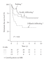
Purpose. Infiltrative microscopical peripheral growth of soft tissue sarcomas (STS) has been shown to be of prognostic importance and preoperative risk stratification could individualize neoadjuvant treatment. Patients and methods. We assessed peripheral tumour growth pattern on preoperative MRI from 78 STS. The findings were correlated to histopathology and to outcome. Results. The MRI-based peripheral tumour growth pattern was classified as pushing in 34 tumours, focally infiltrative in 25, and diffusely infiltrative in 19. All tumours with diffuse infiltration on MRI also showed microscopical infiltration, whereas MRI failed to identify infiltration in two-thirds of the microscopically infiltrative tumours. Diffusely infiltrative growth on MRI gave a 2.5 times increased risk of metastases (P = .01) and a 3.7 times higher risk of local recurrence (P = .02). Discussion. Based on this observation we suggest that MRI evaluation of STS should focus on the peripheral tumour growth pattern since it adds prognostic information of value for decisions on neoadjuvant therapies.
Figures


References
-
- Cormier JN, Pollock RE. Soft tissue sarcomas. CA: A Cancer Journal for Clinicians. 2004;54(2):94–109. - PubMed
-
- Trovik CS, Bauer HC, Alvegard TA, et al. Surgical margins, local recurrence and metastasis in soft tissue sarcomas: 559 surgically-treated patients from the Scandinavian Sarcoma Group Register. European Journal of Cancer. 2000;36(6):710–716. - PubMed
-
- Gustafson P, Akerman M, Alvegard TA, et al. Prognostic information in soft tissue sarcoma using tumour size, vascular invasion and microscopic tumour necrosis-the SIN-system. European Journal of Cancer. 2003;39(11):1568–1576. - PubMed
-
- Wunder JS, Healey JH, Davis AM, Brennan MF. A comparison of staging systems for localized extremity soft tissue sarcoma. Cancer. 2000;88(12):2721–2730. - PubMed
-
- Varma DG. Imaging of soft-tissue sarcomas. Current Oncology Reports. 2000;2(6):487–490. - PubMed
LinkOut - more resources
Full Text Sources
Other Literature Sources

