. 2007;2007(1):25935.
doi: 10.1155/2007/25935. Epub 2007 Feb 28.

Distinction between pore assembly by staphylococcal alpha-toxin versus leukotoxins

Affiliations

Distinction between pore assembly by staphylococcal alpha-toxin versus leukotoxins

Olivier Joubert et al. J Biomed Biotechnol. 2007.

Abstract

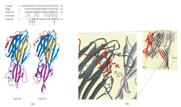
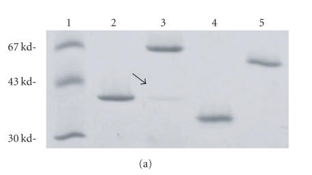
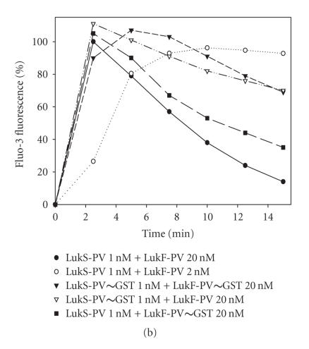
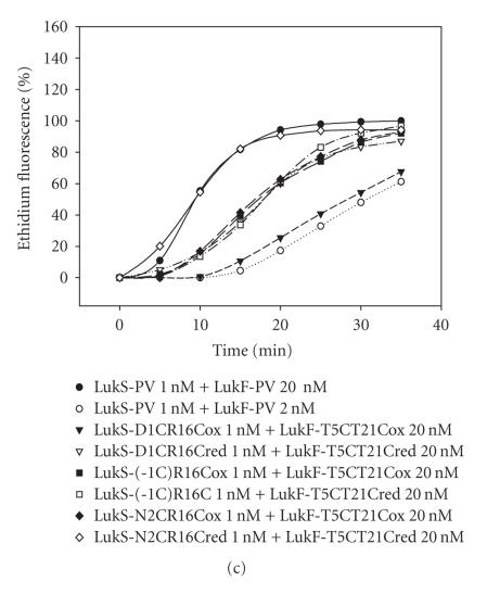
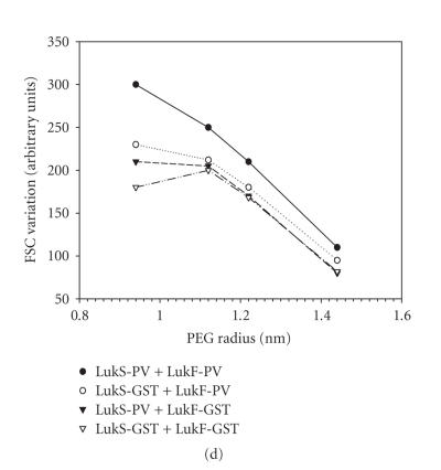
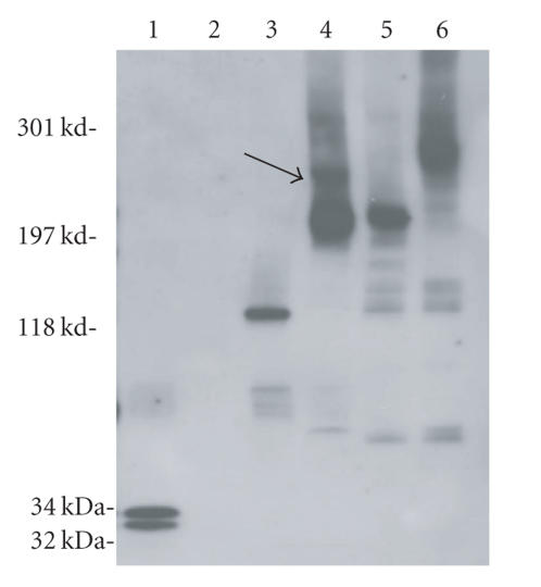
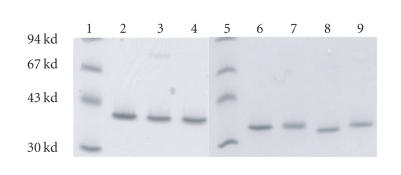
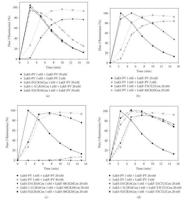
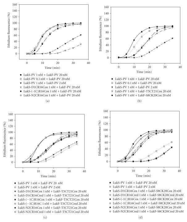
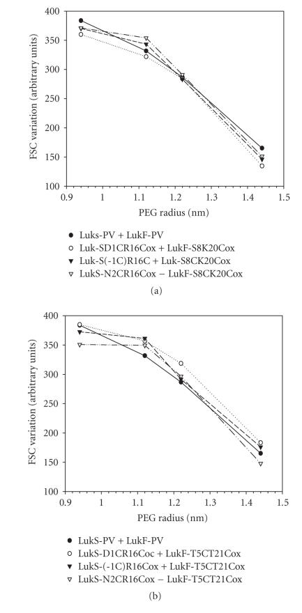
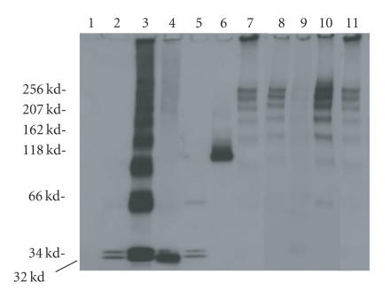
The staphylococcal bipartite leukotoxins and the homoheptameric alpha-toxin belong to the same family of beta-barrel pore-forming toxins despite slight differences. In the alpha-toxin pore, the N-terminal extremity of each protomer interacts as a deployed latch with two consecutive protomers in the vicinity of the pore lumen. N-terminal extremities of leukotoxins as seen in their three-dimensional structures are heterogeneous in length and take part in the beta-sandwich core of soluble monomers. Hence, the interaction of these N-terminal extremities within structures of adjacent monomers is questionable. We show here that modifications of their N-termini by two different processes, using fusion with glutathione S-transferase (GST) and bridging of the N-terminal extremity to the adjacent beta-sheet via disulphide bridges, are not deleterious for biological activity. Therefore, bipartite leukotoxins do not need a large extension of their N-terminal extremities to form functional pores, thus illustrating a microheterogeneity of the structural organizations between bipartite leukotoxins and alpha-toxin.


Figures

Figure 1
Figure 1
Structural features of the N-terminal extremities of staphylococcal bipartite leukotoxins and α-toxin. (a) Sequence alignment of the N-termini of staphylococcal α-toxin and of the two components LukF-PV and LukS-PV of the Panton-Valentine leucocidin. The strands arrangement of LukF-PV is indicated by dashes. Cysteine-substituted residues, indicated in bold in the sequences, are also shown on the 3D structures of LukF-PV (PDB code 1pvl) and LukS-PV (PDB code 1t5r). (b) For comparison, view three protomers of the α-toxin heptamer (PDB code 7AHL) and polar interactions involving the N-terminal extremity of a given subunit (red) with residues of two adjacent protomers (grey and dark grey) within the α-toxin pore lumen.
Figure 2
Figure 2
Biological activity of GST fusion proteins. (a) Control of homogeneity by 3%–8% (w/vol) SDS-PAGE of 200 ng of each purified protein is shown; (1) molecular ladder, (2) LukF-PV, (3) GST∼LukF-PV, (4) LukS-PV, (5) GST∼LukS-PV. (b) Flow cytometry evaluation of the Ca2+ entry into human PMNs mediated by combinations of wild-type LukS-PV and LukF-PV and GST fusion proteins. (c) Flow cytometry evaluation of the ethidium entry into human PMNs mediated by combinations of LukS-PV and LukF-PV and GST fusion proteins. (d) Hydrodynamic radius of pores formed by WT and fusion proteins in human PMNs determined after a 30-minute incubation of toxins (20 nM of S and 100 nM of F components). Osmotic protection by polyethylene glycol molecules was assessed by variations of the mean FSC (forward light scatter) value in the presence of PEG molecules of different hydrodynamic radii.
Figure 2
Figure 2
Biological activity of GST fusion proteins. (a) Control of homogeneity by 3%–8% (w/vol) SDS-PAGE of 200 ng of each purified protein is shown; (1) molecular ladder, (2) LukF-PV, (3) GST∼LukF-PV, (4) LukS-PV, (5) GST∼LukS-PV. (b) Flow cytometry evaluation of the Ca2+ entry into human PMNs mediated by combinations of wild-type LukS-PV and LukF-PV and GST fusion proteins. (c) Flow cytometry evaluation of the ethidium entry into human PMNs mediated by combinations of LukS-PV and LukF-PV and GST fusion proteins. (d) Hydrodynamic radius of pores formed by WT and fusion proteins in human PMNs determined after a 30-minute incubation of toxins (20 nM of S and 100 nM of F components). Osmotic protection by polyethylene glycol molecules was assessed by variations of the mean FSC (forward light scatter) value in the presence of PEG molecules of different hydrodynamic radii.
Figure 3
Figure 3
Oligomers formed by PVL and modified toxins. Oligomers were checked in solution or after recovery from treated human PMNs, 3–8% (w/v) SDS-PAGE and immunoblotting with anti-LukS-PV and anti-LukF-PV affinity-purified rabbit antibodies. Lane 1: LukS-PV + LukF-PV without membranes, lane 2: PMNs only, lane 3: GST∼LukS-PV + GST∼LukF-PV, lanes 4, 5, 6: toxins applied on PMNs membranes and then saponin/glutaraldehyde treated and heated 5 minutes at 100°C as described in materials and methods, lane 4: LukS-PV + GST∼LukF-PV, lane 5: GST∼LukS-PV + LukF-PV, lane 6: GST∼LukS-PV + GST∼LukF-PV.
Figure 4
Figure 4
Control of homogeneity and absence of significant dimerization in nonreducing conditions by 3–8% (w/v) SDS-PAGE of 200 ng of each mutated protein: lane 1: molecular ladder, lane 2: LukF-PV, lane 3: LukF-PV T5C-T21C oxidized (ox), lane 4: LukF-PV S8C-K20Cox, lane 5: molecular ladder, lane 6: LukS-PV, lane 7: LukS-PV-1C-R16Cox, lane 8: LukS-PV D1C-R16Cox, lane 9: LukS-PV N2C-R16Cox.
Figure 5
Figure 5
Flow cytometry evaluation of Ca2+ entry into human PMNs for different combinations of purified WT S and F proteins and their oxidized double mutants. (a) Oxidized LukS-PV mutants were tested with WT LukF-PV. (b) Oxidized LukF-PV double mutants were tested with WT LukS-PV. (c) Oxidized LukS-PV double mutants were tested with LukF-PV S8C-K20Cox. (d) Oxidized LukS-PV double mutants were tested with LukF-PV T5C-T21Cox.
Figure 6
Figure 6
Flow cytometry evaluation of ethidium entry into human PMNs for different combinations of purified WT S and F proteins and their oxidized or reduced double mutants. (a) Oxidized LukS-PV mutants were first tested with WT LukF-PV. (b) Oxidized LukF-PV mutants were tested with WT LukS-PV. (c) Oxidized or reduced LukS-PV double mutants were tested with LukF-PV S8C-K20C. (d) Oxidized or reduced LukS-PV double mutants were tested with LukF-PV T5C-T21C.
Figure 7
Figure 7
Determination of the hydrodynamic radius of pores formed by the different combinations of oxidized LukS-PV (20 nM) and LukF-PV (100 nM) mutants 30 minutes after application of toxins to human PMNs. Osmotic protection was assessed by variations of the mean FSC (forward light scatter) value in the presence of polyethylene glycol molecules of different hydrodynamic radii. (a) Oxidized LukS-PV double mutants were tested with LukF-PV S8C-K20Cox. (b) Oxidized LukS-PV double mutants were tested with LukF-PV T5C-T21Cox.
Figure 8
Figure 8
Oligomers formed by PVL and modified toxins. Oligomers were checked in solution or after recovery from treated human PMNs, 3–8% (w/v) SDS-PAGE and immunoblotting with anti-LukS-PV and anti-LukF-PV affinity-purified rabbit antibodies. Lane 1: PMNs only, lane 2: LukS-PV + LukF-PV in solution (4 ng each), lane 3: LukS-PV + LukF-PV (4 ng each/load) treated by 0.3 mM glutaraldehyde, lane 4: LukS-PV + LukF-PV saponin/treated with 3 mM glutaraldehyde. Lanes 5–11: each PVL components (30 ng each/load) was applied on PMNs, lane 5: LukS-PV + LukF-PV saponin treated, lane 6: LukS-PV + LukF-PV saponin- and glutaraldehyde treated without heating at 100°C, lane 7: LukS-PV + LukF-PV saponin at 0°C/glutaraldehyde treated and boiling at 100°C, lanes 8–11: toxins applied on PMNs, saponin treatment at room temperature + 1 mM glutaraldehyde and boiling, lane 8: LukS-PV + LukF-PV, lane 9: LukS-PV-1C-R16C + LukF-PV S8C-K20C, lane 10: LukS-PV N2C-R16C + LukF-PV S8C-K20C, lane 11: LukS-PV + LukF-PV oxidized.

References

    1. Prévost G. Toxins in Staphylococcus aureus pathogenesis. In: Proft T, editor. Microbial Toxins. Molecular and Cellular Biology. Norfolk, UK: Horizon Bioscience; 2005. pp. 243–284.
    1. Nguyen VT, Kamio Y, Higuchi H. Single-molecule imaging of cooperative assembly of γ-hemolysin on erythrocyte membranes. EMBO Journal. 2003;22(19):4968–4979. - PMC - PubMed
    1. Miles G, Movileanu L, Bayley H. Subunit composition of a bi-component toxin: staphylococcal leukocidin forms an octameric transmembrane pore. Protein Science. 2002;11(4):894–902. - PMC - PubMed
    1. Ferreras M, Höper F, Dalla Serra M, Colin DA, Prévost G, Menestrina G. The interaction of Staphylococcus aureus bi-component γ-hemolysins and leucocidins with cells and lipid membranes. Biochimica et Biophysica Acta (BBA) - Biomembranes. 1998;1414(1-2):108–126. - PubMed
    1. Jayasinghe L, Bayley H. The leukocidin pore: evidence for an octamer with four LukF subunits and four LukS subunits alternating around a central axis. Protein Science. 2005;14(10):2550–2561. - PMC - PubMed