

Localization of human intraparietal areas AIP, CIP, and LIP using surface orientation and saccadic eye movement tasks
- PMID: 17497631
- PMCID: PMC6870972
- DOI: 10.1002/hbm.20396
Localization of human intraparietal areas AIP, CIP, and LIP using surface orientation and saccadic eye movement tasks
Abstract
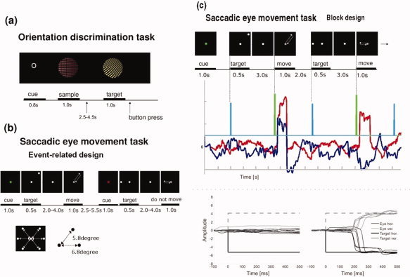
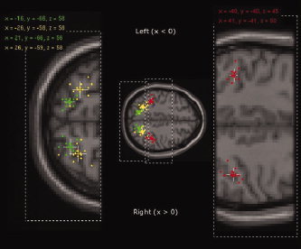
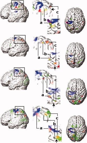
In monkeys, areas in the intraparietal sulcus (IPS) play a crucial role in visuospatial information processing. Despite many human neuroimaging studies, the location of the human functional homologs of some IPS areas is still a matter of debate. The aim of the present functional magnetic resonance imaging (fMRI) study was to identify the distinct locations of specific human IPS areas based on their functional properties using stimuli adapted from nonhuman primate experiments, in particular, surface orientation discrimination and memory guided saccadic eye movements (SEM). Intersubject anatomical variability likely accounts for much of the debate. By applying subject by subject analysis, we can demonstrate that sufficient intersubject anatomical and functional commonalities exist. Both the lateral bank of the anterior part of IPS, the putative human homolog of the area AIP, and the caudal part of the IPS (putative CIP) showed activation related to spatial discrimination of surface orientation. Eye tracking conducted during fMRI data acquisition allowed us to show that both areas were separated by an area related to SEM. This area was located in the middle region of the IPS (most probably including LIP), i.e., similar to the location observed in nonhuman primates. In 10 of 11 subjects our putative CIP activation was located in a medial side branch of the posterior part of the IPS, on the opposite side as described in nonhuman primates, making this landmark a useful anatomical marker for the location of CIP.
(c) 2007 Wiley-Liss, Inc.
Figures
Similar articles
-
Functional properties and interaction of the anterior and posterior intraparietal areas in humans.Eur J Neurosci. 2003 Mar;17(5):1105-10. doi: 10.1046/j.1460-9568.2003.02540.x. Eur J Neurosci. 2003. PMID: 12653987
-
The functional organization of the intraparietal sulcus in humans and monkeys.J Anat. 2005 Jul;207(1):3-17. doi: 10.1111/j.1469-7580.2005.00426.x. J Anat. 2005. PMID: 16011542 Free PMC article. Review.
-
From three-dimensional space vision to prehensile hand movements: the lateral intraparietal area links the area V3A and the anterior intraparietal area in macaques.J Neurosci. 2001 Oct 15;21(20):8174-87. doi: 10.1523/JNEUROSCI.21-20-08174.2001. J Neurosci. 2001. PMID: 11588190 Free PMC article.
-
Differential effects of viewpoint on object-driven activation in dorsal and ventral streams.Neuron. 2002 Aug 15;35(4):793-801. doi: 10.1016/s0896-6273(02)00803-6. Neuron. 2002. PMID: 12194877
-
Human cortical areas underlying the perception of optic flow: brain imaging studies.Int Rev Neurobiol. 2000;44:269-92. doi: 10.1016/s0074-7742(08)60746-1. Int Rev Neurobiol. 2000. PMID: 10605650 Review.
Cited by
-
Analysis of haptic information in the cerebral cortex.J Neurophysiol. 2016 Oct 1;116(4):1795-1806. doi: 10.1152/jn.00546.2015. Epub 2016 Jul 20. J Neurophysiol. 2016. PMID: 27440247 Free PMC article. Review.
-
Disentangling Representations of Object and Grasp Properties in the Human Brain.J Neurosci. 2016 Jul 20;36(29):7648-62. doi: 10.1523/JNEUROSCI.0313-16.2016. J Neurosci. 2016. PMID: 27445143 Free PMC article.
-
Visual-manual exploration and posterior parietal cortex in humans.J Neurophysiol. 2009 Dec;102(6):3433-46. doi: 10.1152/jn.90785.2008. Epub 2009 Oct 7. J Neurophysiol. 2009. PMID: 19812283 Free PMC article.
-
The organization and evolution of dorsal stream multisensory motor pathways in primates.Front Neuroanat. 2011 Jun 13;5:34. doi: 10.3389/fnana.2011.00034. eCollection 2011. Front Neuroanat. 2011. PMID: 21716641 Free PMC article.
-
The representation of tool and non-tool object information in the human intraparietal sulcus.J Neurophysiol. 2013 Jun;109(12):2883-96. doi: 10.1152/jn.00658.2012. Epub 2013 Mar 27. J Neurophysiol. 2013. PMID: 23536716 Free PMC article.
References
-
- Adams DL ( 1997): Functional organization of the monkey visual cortex for stereoscopic depth. PhD. Thesis. London: University College London.
-
- Binkofski F,Dohle C,Posse S,Stephan KM,Hefter H,Seitz RJ,Freund H‐J ( 1998): Human anterior intraparietal area subserves prehension. Neurology 50: 1253–1259. - PubMed
-
- Binkofski F,Buccino G,Posse S,Seitz RJ,Rizzolatti G,Freund H‐J ( 1999): A fronto‐parietal circuit for object manipulation in man: Evidence from an fMRI study. Eur J Neurosci 11: 3276–3286. - PubMed
Publication types
MeSH terms
LinkOut - more resources
Full Text Sources
Miscellaneous
