. 2007 Dec 1;98(5):1071-82.
doi: 10.1002/bit.21486.

A stirred microchamber for oxygen consumption rate measurements with pancreatic islets

Affiliations

A stirred microchamber for oxygen consumption rate measurements with pancreatic islets

Klearchos K Papas et al. Biotechnol Bioeng. .

Abstract

Improvements in pancreatic islet transplantation for treatment of diabetes are hindered by the absence of meaningful islet quality assessment methods. Oxygen consumption rate (OCR) has previously been used to assess the quality of organs and primary tissue for transplantation. In this study, we describe and characterize a stirred microchamber for measuring OCR with small quantities of islets. The device has a titanium body with a chamber volume of about 200 microL and is magnetically stirred and water jacketed for temperature control. Oxygen partial pressure (pO(2)) is measured by fluorescence quenching with a fiber optic probe, and OCR is determined from the linear decrease of pO(2) with time. We demonstrate that measurements can be made rapidly and with high precision. Measurements with betaTC3 cells and islets show that OCR is directly proportional to the number of viable cells in mixtures of live and dead cells and correlate linearly with membrane integrity measurements made with cells that have been cultured for 24 h under various stressful conditions.


Figures
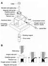
Figure 1
Figure 1
Schematic diagram of device for measuring oxygen consumption. A: Exploded view. The device consists of a titanium cup that sits in a water jacketed enclosure for temperature control. After addition of a glass-coated magnetic stirring bar (nominal length 5 mm, diameter 2 mm) into the opening in the cup, a transparent beveled glass plug is placed into the cup opening (diameter 6.4 mm). The void space remaining in the opening defines the chamber that contains the cell or tissue suspensions. Oxygen in the chamber is measured by fluorescence quenching following oxygen binding to a flourophor in a gel overlain by a silicone rubber film at the tip of an optical fiber. The fiber is held inside the chamber by a titanium jacket and elastomeric seal. A magnet attached to a rotating motor is paced within the enclosure in close proximity to the stirring bar. B: Cross section through the titanium cup showing the filling procedure. After the magnetic stirring bar is added, a volume of the cell or tissue suspension corresponding to the chamber volume plus 5–10 µL excess is placed in the chamber (left figure), the beveled plug is inserted, and any excess fluid is expelled through the angled side port and collected in a groone around the cap. When filling is complete, the beveled plug is rotated (right figure) to block access to the port and seal the chamber. If a bubble is observed from the top, the plug is removed, cells or islets are allowed to settle, additional medium is added, and the plug is reinserted as the bubble is washed through the angled side port.
Figure 2
Figure 2
Actual traces (individual data points) of measured pO2 versus time with rat islet samples. A: OCR measurements, each about 20–50 min with a stirring rotational rate of 53 rpm, were performed with a fresh aliquot obtained from the same islet preparation. The measured cell concentration of the islets in the chamber was 1.6 × 10⁶ cells/mL, corresponding to an islet concentration of 1030 IE/mL. Data were fitted to a straight line in the steepest portion of the trace (indicated by vertical marks), yielding slopes listed n the figure. Points below a pO2 of 60 mmHg were not used in any of the slope estimates to ensure that all cells within the islets were exposed to a high enough pO2 so that OCR could be assumed constant throughout the islet. If the experiment is run long enough to allow pO2 to decrease to much lower values, curvature occurs in the plot of pO2 versus time, as shown in the third measurements, which reflects the interaction of intra-islet oxygen gradients and the decrease in OCR as the local pO2 approaches 0 mmHg. B: Two measurements performed with the same sample, the second one after re-oxygenation, with a stirrer rotational rate of 53 rpm and 1.7 × 10⁶ islet cells/mL, corresponding to 1090 IE/mL. The mean of the slopes was 1.77 mmHg/min, corresponding to an OCR of 4.75 nmol/min and an OCR/cell of 1.33 fmol/min · cell. C: Sequential OCR measurements made 4–6 h after isolation with different samples from the same rat islet preparation at stirring speed settings of, “9,” “3,” and “6”, corresponding to rotational rates of 321, 53, and 187 rpm, respectively. D: Trace of each experiment shown in (C) with time adjusted so that pO2 = 0 at t = 0. E: pO2 versus adjusted time from OCR measurements performed with rat islets made within 10 min after isolation was completed at three stirring speed settings. F: Data from OCR measurement performed with islets from same preparation as used in (E) but made 4 h later.
Figure 3
Figure 3
Precision of the OCR measurement. Coefficient of variation (COV) versus the OCR measured in the experiment (bottom) or the corresponding estimated number of viable islet equivalents (top) for measurements conducted with rat, porcine, and human islets in the OCR measurement apparatus. Data for single and triplicate measurements are shown. For illustrative purposes, the number of viable islet equivalents was calculated assuming (OCR/DNA)viable = 500 nmol/min · mg DNA, 6.5 pg DNA/cell, and 1,560 cells/IE (Pisania, 2007a).
Figure 4
Figure 4
Dependence of OCR on cell viability. In (A, B, and C), viability was assessed by membrane integrity measurements with trypan blue. A: OCR as a function of the number of viable cells and (B) OCR/cell as a function of mixture composition for healthy, heat-treated, and mixtures of healthy and heat-treated βTC3 cells in ratios of 25/75, 50/50, and 75/25. Heat-killed cells were incubated at 60°C for 1 h. C: OCR and viable cell number from various batches of βTC3 cells cultured for 24 h with and without imposed stressful conditions. Aliquots for membrane integrity and corresponding OCR measurement were taken from the same sample. The solid line is the best fit of a line through the origin by linear regression (R2 = 0.983). The estimate of the slope was 1.72 ± 0.03 fmol/min · viable cell. D: Relationship between MTT absorbance and viable cell number. βTC3 cells from a single batch were cultured for 24 h under unstressed (control) or stressed conditions (TNF-α, 500 U/mL culture medium). E: MTT absorbance versus OCR for porcine islets from a single preparation cultured under normal and stressful conditions. Islets were cultured for 24 h in multi-well plates at 37°C with medium depths of 3 and 10 mm and with fractional islet surface coverage ranging from 0.4% (control) to 30%. At the end of the incubation period islets were removed from the corresponding wells for MTT and OCR measurements. F: MTT versus OCR for samples of roughly similar islet volume from 17 different rat islets preparations (R2 = 0.993). In (A) through (F), measurements were made in triplicate. Error bars indicate standard deviation and are contained within domain of symbol if not visible.
Figure 5
Figure 5
Fraction of βTC3 cells that were membrane-impermeable after culture under normal and a variety of stressful conditions for 24 h. Approximately 2 × 10⁷ βTC3 cells were used in each experiment. For surface-attached cultures, cells were cultured in T-75 flasks in a 37°C incubator at a plating density such that they would not be confluent after 24 h. Medium depth was 2 mm. For pelletized culture, cells were placed in 15 mL centrifuge tubes with 15 mL culture media and let settle by gravity, leading to cell depth of about 3 mm overlain by 11 cm of media. The tubes were sealed and cultured at temperatures of 5, 24, and 37°C. For hyperoxic and anoxic culture, flasks, and tubes were placed in sealed chambers within an incubator, and the sealed chambers were continuously flushed with premixed gases (95% O2 or 95% N2, balance CO2). At the end of the 24-h culture, cells in flasks were trypsinized. All cells from each condition were collected and samples for OCR and trypan blue staining were taken. Fraction of membrane-impermeable cells was determined as the number of cells that did not take up trypan blue divided by the total number of cells counted.
Figure 6
Figure 6
Frequency distribution of OCR per viable cell for stressed and unstressed βTC3 cells cultured for 24 h under stressed and unstressed conditions (Figures 4C and 5). OCR per viable cell was estimated by dividing the measured OCR by the number of viable cells placed in the chamber at the time of the measurement as determined with a hemocytometer using trypan blue exclusion. Values of OCR/viable cell ranged between 1.46 and 2.81 (n = 33) for stressed and 0.94–2.99 fmol/min · viable cell (n = 31) for unstressed cells. Mean values were 1.88 ± 0.54 and 1.84 ± 0.34 fmol/min · viable cell (n = 31) for stressed and unstressed cells, respectively. Fractional viability by trypan blue exclusion averaged 0.95, and the OCR uncorrected for viability was 1.75 ± 0.32 fmol/min · cell. OCR values for stressed cells covered a wider range, but the mean values of OCR for stressed and unstressed cells were not significantly different. The mean value for all measurements was 1.86 ± 0.45 (n = 64).

Similar articles

Cited by

References

    1. Arain S, Svenja W, Heinzle E, Gernot JT, Krause C, Klimant I. Sensing microplates with optodes: Influence of oxygen exchange between sample, air, and plate material. Biotechnol Bioeng. 1998;90:271–280. - PubMed
    1. Avgoustiniatos ES. Ph.D. Thesis. Cambridge: Massachusetts Institute of Technology; 2001. Oxygen diffusion limitation in pancreatic islet culture and immunoisolation.
    1. Bank HL. Assessment of islet cell viability using fluorescent dyes. Diabetologia. 1987;30:812–816. - PubMed
    1. Bank HL. Rapid assessment of islet viability with acridine orange and propidium iodide. In Vitro Cell Dev Biol. 1988;24:266–273. - PubMed
    1. Berridge MV, Tan AS. Characterization of the cellular reduction of 3-(4,5-dimethylthiazol-2- yl)-2,5-diphenyltetrazolium bromide (MTT): Subcellular localization, substrate dependence, and involvement of mitochondrial electron transport in MTT reduction. Arch Biochem Biophys. 1993;303:474–482. - PubMed

Publication types

MeSH terms

LinkOut - more resources