. 2007 Jun 5;46(22):6536-46.
doi: 10.1021/bi6026506. Epub 2007 May 12.

Biochemical and crystallographic analysis of substrate binding and conformational changes in acetyl-CoA synthetase

Affiliations

Biochemical and crystallographic analysis of substrate binding and conformational changes in acetyl-CoA synthetase

Albert S Reger et al. Biochemistry. .

Abstract

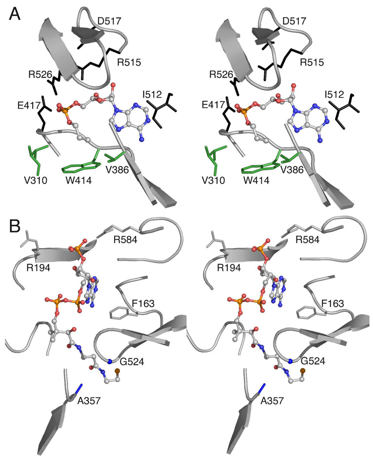
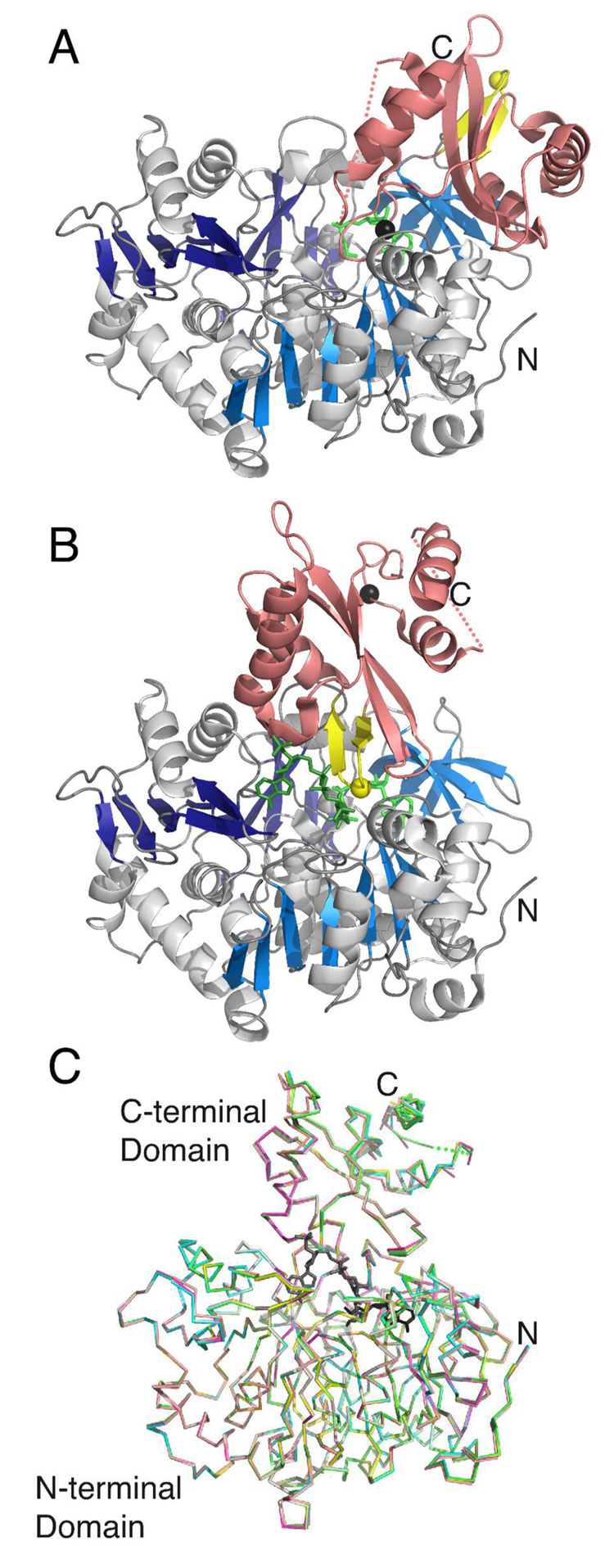
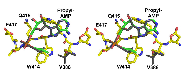
The adenylate-forming enzymes, including acyl-CoA synthetases, the adenylation domains of non-ribosomal peptide synthetases (NRPS), and firefly luciferase, perform two half-reactions in a ping-pong mechanism. We have proposed a domain alternation mechanism for these enzymes whereby, upon completion of the initial adenylation reaction, the C-terminal domain of these enzymes undergoes a 140 degrees rotation to perform the second thioester-forming half-reaction. Structural and kinetic data of mutant enzymes support this hypothesis. We present here mutations to Salmonella enterica acetyl-CoA synthetase (Acs) and test the ability of the enzymes to catalyze the complete reaction and the adenylation half-reaction. Substitution of Lys609 with alanine results in an enzyme that is unable to catalyze the adenylate reaction, while the Gly524 to leucine substitution is unable to catalyze the complete reaction yet catalyzes the adenylation half-reaction with activity comparable to the wild-type enzyme. The positions of these two residues, which are located on the mobile C-terminal domain, strongly support the domain alternation hypothesis. We also present steady-state kinetic data of putative substrate-binding residues and demonstrate that no single residue plays a dominant role in dictating CoA binding. We have also created two mutations in the active site to alter the acyl substrate specificity. Finally, the crystallographic structures of wild-type Acs and mutants R194A, R584A, R584E, K609A, and V386A are presented to support the biochemical analysis.


Figures

Figure 1
Figure 1. Active site interactions of Acs
The structure of the wild-type Acs enzyme determined in the presence of propyl-AMP and CoA (1PG4) and amino acids that form the active site pocket are shown. A. The orientation of propyl-AMP bound to Acs in the thioester-forming conformation illustrates residues that interact that interact with the nucleotide. Interactions made to the nucleotide include Arg526 and Arg515, which interact with the phosphate group. The acetate binding pocket is formed by the three side chains of Val310, Val386, and Trp414 (shown in green). B. The CoA binding site. The nucleotide of CoA binds at the surface of the protein and the pantetheine group leads in to the adenylate site through a tunnel that is bordered by Ala357 and Gly524, two residues that were mutated to contain bulkier side chains. Arg584 was positioned to interact with the CoA 3' phosphate. Arg194 was oriented away from the phosphate but could interact through a torsional rotation.
Figure 2
Figure 2. Ligand density for the wild-type and mutant structures
Omit maps were generated by removing the ligands followed by a cycle of refinement. The maps of the coefficients Fo-Fc were contoured at 2.5σ and shown for the CoA ligand of wild-type (A), V386A (B), and K609A (C). Maps were generated in the same way for the propyl-AMP inhibitor of the R194A (D), R584A (E), and R584E (F) mutants as well. Neighboring protein atoms, including Arg191, which interacts with the 5' diphosphate of CoA, are labeled in panels (A) and (D).
Figure 3
Figure 3. Three dimensional structure of Acs and the proposed conformational change
Acs is shown in the A. adenylate-forming conformation bound to propyl-AMP and B. thioester-forming conformation bound to CoA and propyl-AMP. The N-terminal domains are shown with blue strands while the C-terminal domains is colored pink. The A8 region of the C-terminal domain is shown in yellow, with Gly524 depicted as a single yellow sphere. The Cα position of Lys609 from the A10 region is shown as a black sphere. While the thioester-forming conformation shown in B represents a crystallographically observed structure for Acs (1PG4), the adenylate-forming conformation represents a computational model that was produced by rotating the C-terminal domain of the Acs protein onto the C-terminal domain of the CBAL structure (1T5D), which was determined bound to 4CBA. The N- and C-terminal residues are labeled N and C, and the gap around residues 623–632 is shown with a dashed line. C. The Superposition of the Cα chain of the five mutant enzymes and wild-type Acs bound to acetate, AMP and CoA. The ligands from the wild-type enzyme are shown in black, while the wild-type protein is depicted with green. The mutant proteins are represented in the following colors: K609A (teal), R194A (magenta), R584A (yellow), R584E (grey), and V386A (peach).
Figure 4
Figure 4. Superposition of the acyl binding pockets of wild-type and V386A mutants
The models were superimposed using the Cα positions of all residues. Atoms from the V386A model are colored blue (nitrogens), red (oxygens) and yellow (protein carbons) or green (ligand carbons). The propyl-AMP ligand and Val386 from the wild-type structure are shown in dark grey and illustrate the occluded orientation torsional angles of the propyl group.
Scheme 1
Scheme 1

References

    1. Sieber SA, Marahiel MA. Molecular mechanisms underlying nonribosomal peptide synthesis: approaches to new antibiotics. Chem Rev. 2005;105:715–738. - PubMed
    1. Marahiel MA, Stachelhaus T, Mootz HD. Modular Peptide Synthetases Involved in Nonribosomal Peptide Synthesis. Chem Rev. 1997;97:2651–2674. - PubMed
    1. Challis GL, Naismith JH. Structural aspects of non-ribosomal peptide biosynthesis. Curr Opin Struct Biol. 2004;14:748–756. - PMC - PubMed
    1. Babbitt PC, Kenyon GL, Martin BM, Charest H, Slyvestre M, Scholten JD, Chang KH, Liang PH, Dunaway-Mariano D. Ancestry of the 4-chlorobenzoate dehalogenase: analysis of amino acid sequence identities among families of acyl:adenyl ligases, enoyl-CoA hydratases/isomerases, and acyl-CoA thioesterases. Biochemistry. 1992;31:5594–5604. - PubMed
    1. Dunaway-Mariano D, Babbitt PC. On the origins and functions of the enzymes of the 4-chlorobenzoate to 4-hydroxybenzoate converting pathway. Biodegradation. 1994;5:259–276. - PubMed

Publication types

MeSH terms

LinkOut - more resources