. 2007 May 11:7:74.
doi: 10.1186/1471-2148-7-74.

Evolution of pigment synthesis pathways by gene and genome duplication in fish

Affiliations

Evolution of pigment synthesis pathways by gene and genome duplication in fish

Ingo Braasch et al. BMC Evol Biol. .

Abstract

Background: Coloration and color patterning belong to the most diverse phenotypic traits in animals. Particularly, teleost fishes possess more pigment cell types than any other group of vertebrates. As the result of an ancient fish-specific genome duplication (FSGD), teleost genomes might contain more copies of genes involved in pigment cell development than tetrapods. No systematic genomic inventory allowing to test this hypothesis has been drawn up so far for pigmentation genes in fish, and almost nothing is known about the evolution of these genes in different fish lineages.

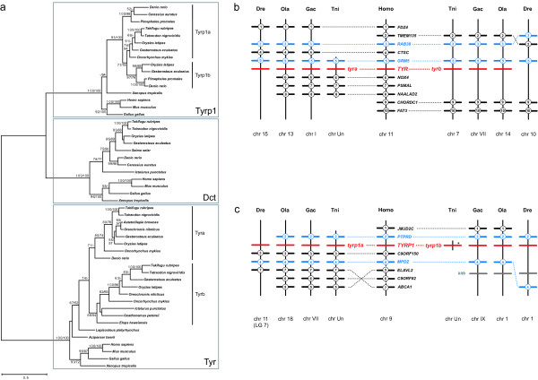
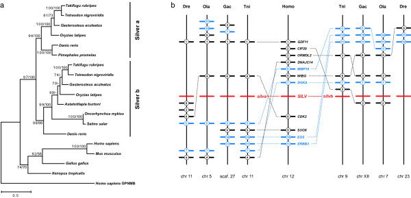
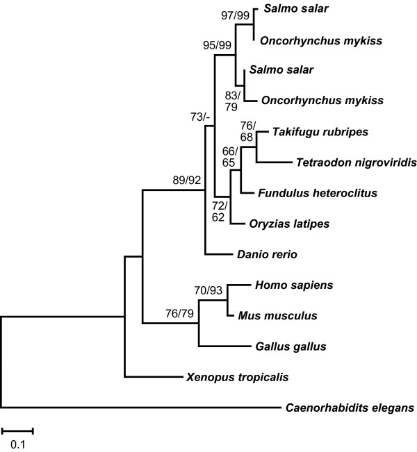
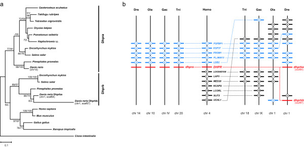
Results: Using a comparative genomic approach including phylogenetic reconstructions and synteny analyses, we have studied two major pigment synthesis pathways in teleost fish, the melanin and the pteridine pathways, with respect to different types of gene duplication. Genes encoding three of the four enzymes involved in the synthesis of melanin from tyrosine have been retained as duplicates after the FSGD. In the pteridine pathway, two cases of duplicated genes originating from the FSGD as well as several lineage-specific gene duplications were observed. In both pathways, genes encoding the rate-limiting enzymes, tyrosinase and GTP-cyclohydrolase I (GchI), have additional paralogs in teleosts compared to tetrapods, which have been generated by different modes of duplication. We have also observed a previously unrecognized diversity of gchI genes in vertebrates. In addition, we have found evidence for divergent resolution of duplicated pigmentation genes, i.e., differential gene loss in divergent teleost lineages, particularly in the tyrosinase gene family.

Conclusion: Mainly due to the FSGD, teleost fishes apparently have a greater repertoire of pigment synthesis genes than any other vertebrate group. Our results support an important role of the FSGD and other types of duplication in the evolution of pigmentation in fish.


Figures

Figure 1
Figure 1
Eumelanin synthesis pathway and gene duplications in vertebrates. Eumelanin is synthesized from tyrosine within the melanosome of melanophores. This requires members of the Tyrosinase family (TYR, DCT, TYRP1) and probably Silver (SILV). Three melanosomal transporters (OCA2, AIM1 and SLC24A5) are crucial for proper melanin synthesis. Red indicates duplications during the fish-specific genome duplication.
Figure 2
Figure 2
Evolution of the tyrosinase gene family in vertebrates. (a) Maximum-likelihood phylogeny of protein sequences from the tyrosinase family based on 570 AA positions. The tree is mid-point rooted. Numbers at the branches denote bootstrap values (maximum likelihood/neighbor joining). Bootstrap values above 50 are shown. Tyrp1a and Tyrp1b are assigned according to the analysis of their genomic environment. (b) Synteny of tyr-containing regions in vertebrate genomes. The human TYR region is syntenic to two tyr paralogons in Tetraodon (Tni), stickleback (Gac) and medaka (Ola). Tyrb was apparently lost in the zebrafish (Dre). (c) Synteny of tyrp1-containing regions in vertebrate genomes. The human TYRP1 region is syntenic to two tyrp1 paralogons in stickleback, medaka and zebrafish. A tyrp1b pseudogene is found in Tetraodon (asterisk). Numbered bars represent genes contributing to conserved synteny, genes that do not contribute to conserved synteny are not shown. Blue bars indicate genes that are duplicated along with tyr or tyrp1b, respectively. Dotted lines connect orthologous genes. Kitb (grey bars), another teleost-specific pigmentation gene duplicate [17] is found 3' of tyrp1b in the four teleost genomes but belongs to a different paralogon (see text).
Figure 3
Figure 3
Evolution of the silver genes in vertebrates. (a) Maximum-likelihood phylogeny of Silver protein sequences based on 523 AA positions. The repeat region [21] was excluded from the alignment. The tree was rooted with human GPNMB. Numbers at the branches denote bootstrap values (maximum likelihood/neighbor joining) above 50%. (b) Synteny of silv-containing regions in vertebrate genomes. The human SILV region is syntenic to two silv paralogons in Tetraodon (Tni), stickleback (Gac), medaka (Ola) and zebrafish (Dre). Numbered bars represent genes contributing to conserved synteny, genes that do not contribute to conserved synteny are not shown. Blue bars indicate genes that are duplicated along with silv. Dotted lines connect orthologous genes.
Figure 4
Figure 4
Pteridine synthesis pathway and gene duplications in vertebrates. Pteridine synthesis contains three component pathways [25]: the de novo synthesis of H4biopterin from GTP (top line), the H4biopterin regeneration pathway (grey) and the synthesis of yellow pteridine pigments. The formation of orange drosopterin has not been elucidated yet in vertebrates. In Drosophila, the clot enzyme is involved [53], which corresponds to the vertebrate Txnl5 protein. Asterisks indicate hypothetical reactions and question marks unidentified enzymes. Red indicates duplications during the fish-specific genome duplication, blue other types of duplication.
Figure 5
Figure 5
Evolution of the GTP-cyclohydrolase I gene family in vertebrates. (a) Maximum-likelihood phylogeny of GchI protein sequences based on 268 AA positions (left). The tree is rooted with GchI from urochordates. Numbers at the branches denote bootstrap values (maximum likelihood/neighbor joining) above 50%. Groups (GchIa, GchIb, GchIc) were assigned according to genomic environment of gchI genes (right). GchIa and gchIb are both linked to members of the socs gene family (blue). GchIb is absent from mammalian and avian genomes, gchIc is only found in some teleost lineages. Dotted lines connect orthologous genes. (b) The gchIb region of teleosts and amphibian is syntenic to a chromosomal block in the genomes of mammals and bird lacking gchIb, suggesting that gchIb was lost secondarily in these lineages.
Figure 6
Figure 6
Molecular phylogeny of the GchI feedback regulatory protein in vertebrates. Maximum-likelihood phylogeny of Gchfr protein sequences based on 95 AA positions. The tree is rooted with Gchfr from nematode. Numbers at the branches denote bootstrap values (maximum likelihood/neighbor joining) above 50%. Gchfr is duplicated in salmon and rainbow trout due to the salmonid-specific tetraploidization.
Figure 7
Figure 7
Evolution of sepiapterin reductase genes in vertebrates. (a) Maximum-likelihood phylogeny of Spr protein sequences based on 313 AA positions. The tree is rooted with Spr from fruitfly. Numbers at the branches denote bootstrap values (maximum likelihood/neighbor joining) above 50%. Groups are assigned according to synteny. (b) Synteny of spr regions in vertebrates. The human SPR region is syntenic to two spr paralogons in Takifugu (Tru), stickleback (Gac) and zebrafish (Dre). sprb was possibly lost in Tetraodon (Tni) and medaka (Ola). Numbered bars represent genes contributing to conserved synteny, genes that do not contribute to conserved synteny are not shown. Blue indicates genes that are duplicated along with spr. Dotted lines connect orthologous genes.
Figure 8
Figure 8
Evolution of pcbd genes in vertebrates. (a) Maximum-likelihood phylogeny of Pcbd proteins based on 107 AA positions. The tree is rooted with Pcbd from sea urchin. Numbers at the branches denote bootstrap values (maximum likelihood/neighbor joining) above 50%. Pcbd was duplicated in vertebrates (Pcbd1 and Pcbd2). In Takifugu, two pcbd1 are observed in scaffolds 53 and 178. The latter (red) is a retro-pseudogene. (b) Exon-intron structure of Pcbd1. Pcbd1 from human and Takifugu scaffold 53 consists of four exons indicated by 4 blocks. Takifugu scaffold 178 contains a "processed" pseudogene with a single exon (bottom row) and a premature stop codon (arrowhead).
Figure 9
Figure 9
Evolution of dihydropteridine reductase genes in vertebrates. (a) Maximum-likelihood phylogeny of Dhpr protein sequences based on 247 AA positions. The tree is rooted with Dhpr from urochordates. Numbers at the branches denote bootstrap values (maximum likelihood/neighbor joining) above 50%. (b) Synteny of dhpr regions in vertebrates. The human DHPR region is syntenic to two paralogons in Tetradon (Tni), stickleback (Gac), medaka (Ola) and zebrafish (Dre). Dhprb was apparently lost in Tetraodon (Tni), stickleback (Gac) and medaka (Ola) and further duplicated in zebrafish, so that two duplicates, dhprba and dhprbb, are found on chromosome 1. Numbered bars represent genes contributing to conserved synteny, genes that do not contribute to conserved synteny are not shown. Blue bars indicate genes that are also duplicated. Dotted lines connect orthologous genes.

References

    1. Fujii R. Coloration and chromatophores. In: Evans DH, editor. The Physiology of Fishes. Boca Raton, Florida: CRC Press; 1993. pp. 535–562.
    1. Bagnara JT. Comparative anatomy and physiology of pigment cells in nonmammalian tissues. In: Nordlund JJ, Boissy RE, Hearing VJ, King RA, Ortonne JP, editor. The Pigmentary System: Physiology and Pathophysiology. New York: Oxford University Press; 1998. pp. 9–40.
    1. Mellgren EM, Johnson SL. The evolution of morphological complexity in zebrafish stripes. Trends Genet. 2002;18:128–34. doi: 10.1016/S0168-9525(01)02614-2. - DOI - PubMed
    1. Odenthal J, Rossnagel K, Haffter P, Kelsh RN, Vogelsang E, Brand M, van Eeden FJ, Furutani-Seiki M, Granato M, Hammerschmidt M, Heisenberg CP, Jiang YJ, Kane DA, Mullins MC, Nusslein-Volhard C. Mutations affecting xanthophore pigmentation in the zebrafish, Danio rerio. Development. 1996;123:391–8. - PubMed
    1. Dutton KA, Pauliny A, Lopes SS, Elworthy S, Carney TJ, Rauch J, Geisler R, Haffter P, Kelsh RN. Zebrafish colourless encodes sox10 and specifies non-ectomesenchymal neural crest fates. Development. 2001;128:4113–25. - PubMed

Publication types