. 2007 Jun 29;370(1):39-52.
doi: 10.1016/j.jmb.2007.04.011. Epub 2007 Apr 12.

Structure of the cytochrome b6f complex: quinone analogue inhibitors as ligands of heme cn

Affiliations

Structure of the cytochrome b6f complex: quinone analogue inhibitors as ligands of heme cn

E Yamashita et al. J Mol Biol. .

Abstract

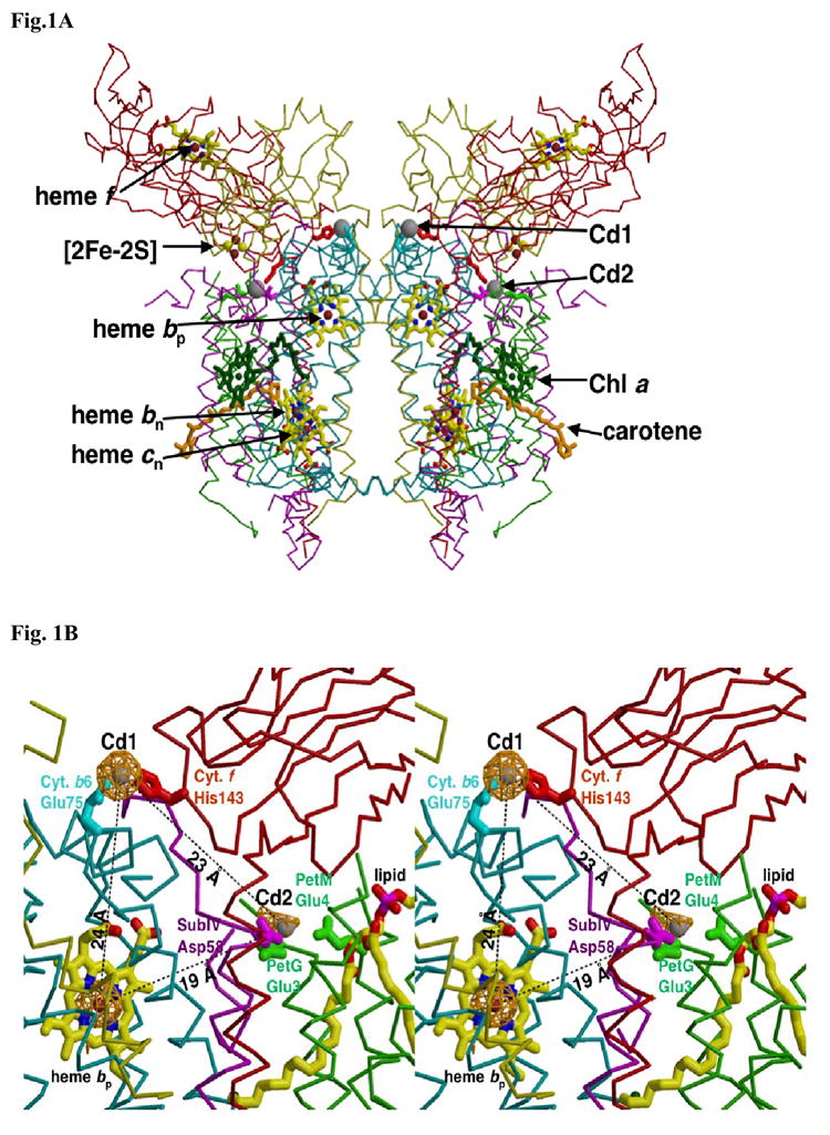
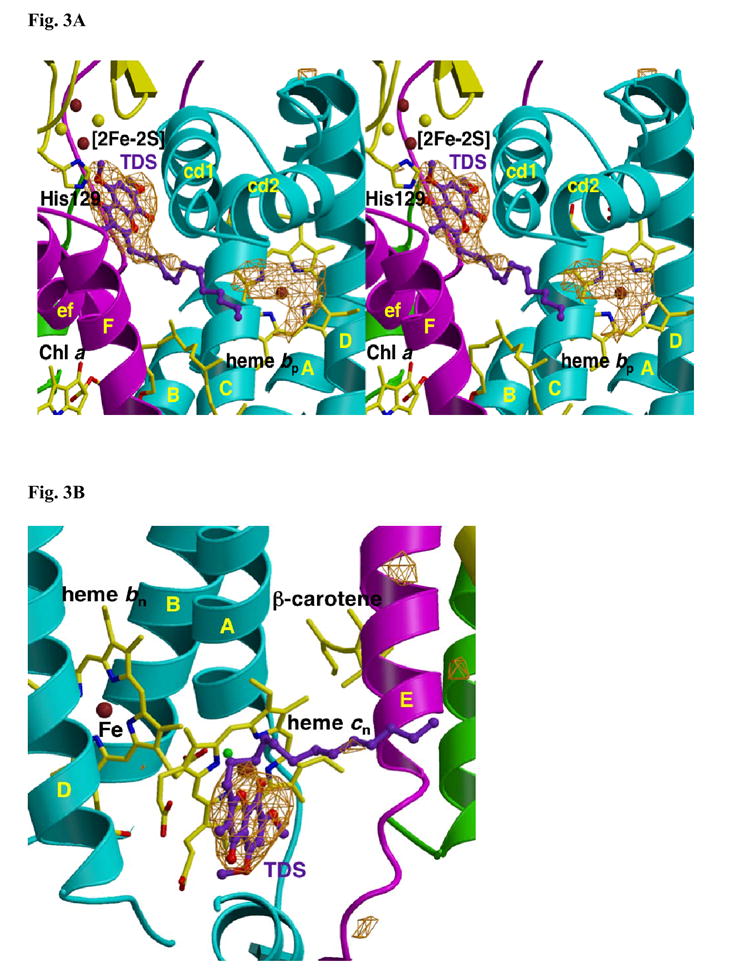
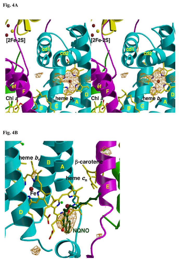
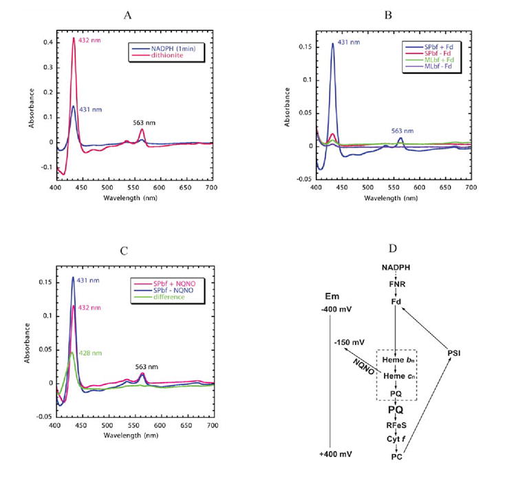
A native structure of the cytochrome b(6)f complex with improved resolution was obtained from crystals of the complex grown in the presence of divalent cadmium. Two Cd(2+) binding sites with different occupancy were determined: (i) a higher affinity site, Cd1, which bridges His143 of cytochrome f and the acidic residue, Glu75, of cyt b(6); in addition, Cd1 is coordinated by 1-2 H(2)O or 1-2 Cl(-); (ii) a second site, Cd2, of lower affinity for which three identified ligands are Asp58 (subunit IV), Glu3 (PetG subunit) and Glu4 (PetM subunit). Binding sites of quinone analogue inhibitors were sought to map the pathway of transfer of the lipophilic quinone across the b(6)f complex and to define the function of the novel heme c(n). Two sites were found for the chromone ring of the tridecyl-stigmatellin (TDS) quinone analogue inhibitor, one near the p-side [2Fe-2S] cluster. A second TDS site was found on the n-side of the complex facing the quinone exchange cavity as an axial ligand of heme c(n). A similar binding site proximal to heme c(n) was found for the n-side inhibitor, NQNO. Binding of these inhibitors required their addition to the complex before lipid used to facilitate crystallization. The similar binding of NQNO and TDS as axial ligands to heme c(n) implies that this heme utilizes plastoquinone as a natural ligand, thus defining an electron transfer complex consisting of hemes b(n), c(n), and PQ, and the pathway of n-side reduction of the PQ pool. The NQNO binding site explains several effects associated with its inhibitory action: the negative shift in heme c(n) midpoint potential, the increased amplitude of light-induced heme b(n) reduction, and an altered EPR spectrum attributed to interaction between hemes c(n) and b(n). A decreased extent of heme c(n) reduction by reduced ferredoxin in the presence of NQNO allows observation of the heme c(n) Soret band in a chemical difference spectrum.


Figures

Fig. 1
Fig. 1. Two cadmium (Cd2+) binding sites on the p-side of the M. laminosus b6f complex
(A) Position of higher occupancy (Cd1) is close to the inter-monomer interface, and that of lower occupancy (Cd2) site is near the small subunits and the exterior of the complex. View is parallel to the plane of the membrane. Distances: (i) from Cd1 site, and (ii) from Cd2, to the [2Fe-2S] cluster on the same and opposite side monomer, (i) 38.9 Å and 40.1 Å, and (ii) 57.1 and 28.0 Å. Color code: cytochrome b6 (cyan), subunit IV (purple), cytochrome f (red), ISP (yellow), PetG, PetL, PetM, and PetN (green). (B, stereo) Environment of Cd1 and Cd2 sites shown in more detail. Lower occupancy of the Cd2 site is shown by the smaller cage of electron density. A lipid molecule (possibly galactolipid) described in the coordinates of the C. reinhardtii b6f complex (pdb; 1Q90), but not previously discussed, is closer to Cd2. Distances: higher (Cd1) to lower occupancy (Cd2) Cd2+ site, 23.2 Å; higher occupancy Cd1 site to heme bp, 24.0 Å; Cd2 to residues Glu78, Trp79 and Y80 of the ‘PEWY’ loop on the same monomer, 13, 12, and 16 Å. The seven peaks of largest amplitude in the anomalous difference map are: Cd1 site (18.5 σ), heme bp (18.1 σ), heme cn (17.6 σ), heme bn (17.0 σ), heme f (13.3 σ), [2Fe-2S] site (11.1 σ), Cd2 site (6.3 σ). The anomalous scattering factors (f″) for Fe and Cd at 0.98 A are 1.50 and 2.13, respectively. The B factors of the Cd1 and Cd2 sites are 80 Å2 and 178 Å2.
Fig. 2
Fig. 2
Fo-Fc difference map on [A] p- and [B] n-side of the native b6f complex in the absence of added inhibitor (“native” structure). TM helices and surface helices within loops are labeled in upper and lower case, respectively. [A] Fo-Fc difference map, contoured at 4σ, in the native (with Cd2+) b6f complex. Origin of electron density under the cd2 loop, presumably lipid and/or detergent, is not known. [B] Fo-Fc difference map of n-side background electron density in the native (with Cd2+) b6f complex. Fo-Fc map contoured at 4σ.
Fig. 3
Fig. 3. Fo-Fc difference map of (A) p- and (B) n-side binding site of TDS in the b6f complex
(A) p-side. The extra density within H-bond distance of the His129 ligand of the [2Fe-2S] cluster and between the ‘ef’ and ‘cd1’ loops is attributed to the chromone ring of TDS. As in Fig. 2A, the origin of the electron density under the cd2 loop, presumably lipid and/or detergent, is not known. (B) n-side. The position of TDS is shown relative to heme cn, which is exposed to the quinone-exchange cavity. TDS is on the side of heme cn distal to heme bn. TM helices and surface helices within loops labeled as in Fig. 2. Fo-Fc maps contoured at 4σ.
Fig. 4
Fig. 4. Fo-Fc difference map of (a) p-side and (b) n-side binding site of NQNO in the b6f complex
(A) As in Fig. 2A, the origin of the electron density under the cd2 loop, presumably lipid and/or detergent, is not known. The p-side plastoquinone binding niche that includes the [2Fe-2S] cluster and the p-side portal in the “roof” of the cavity is shown. (B) The Fo – Fc difference map in the region of heme cn on the n-side of the b6f complex shows the n-side binding site of NQNO. TM helices and surface helices within loops are labeled as in Fig. 2. Fo-Fc maps are contoured at 4σ.
Fig. 5
Fig. 5. Reduction of hemes bn/cn by reduced ferredoxin/FNR
The reaction solution contained 1.5 μM spinach b6f complex with bound FNR and 3 μM spinach ferredoxin. Semi-anaerobic conditions were obtained through the addition of 10 mM glucose and 160 units of glucose oxidase. Decyl-plastoquinol (50 μM) was present to eliminate any contribution of cytochrome f to the difference spectra. Cytochrome b6 /cn reduction was initiated by addition of 0.3 mM NADPH. (A) Comparison of reduction of spinach b6f complex by NADPH/ferredoxin and dithionite. The NADPH/ferredoxin minus decyl-plastoquinol difference spectrum (blue trace) was measured 1 min after addition of NADPH. The spectrum of fully reduced cytochrome b6 (pink) was obtained by addition of dithionite. (B) Dependence of reduction of hemes bn/cn reduction on the presence of FNR and ferredoxin. Reduction of spinach b6f complex following addition of NADPH in the presence (blue) or absence (red) of ferredoxin (3 μM), and of M. laminosus b6f complex, which does not have bound FNR, in the presence (green) or absence (purple) of ferredoxin. All spectra were measured 1 min after addition of NADPH. (C) Double difference spectrum, normalized at 563 nm, showing the Soret band spectrum of heme cn (green), measured as the difference between the spectrum obtained in the presence and absence of NQNO. Difference spectra obtained in the absence (blue) and presence (pink) of NQNO (final concentration, 25 μM). (D) Proposed pathway of reduction of hemes bn and cn by reduced ferredoxin (Fd) in a Q cycle or cyclic pathway in which ferredoxin is reduced physiologically by the photosystem I reaction center and by NADPH in the present experiment. The requirement of FNR and Fd for the pathway of heme bn reduction by NADPH is defined by the data shown in Fig. 5B. The ability of NQNO to block the pathway at the site of heme cn is ascribed to NQNO binding as a ligand of this heme (Fig. 4B) and the NQNO-induced decrease in Em7 of heme cn . The inference that hemes bn and cn, together with PQ, are part of a complex in which PQ serves as a ligand of heme cn is based on the proximity of the two hemes ; , their interaction measured by EPR , and binding of the quinone analogue inhibitors, TDS (Fig. 3B) and NQNO (Fig. 4B).

Similar articles

Cited by

References

    1. Kallas T. The cytochrome b6f complex. In: Bryant DA, editor. The Molecular Biology of Cyanobacteria. Kluwer Academic Publishers; Dordrecht: 1994. pp. 259–317.
    1. O’Keefe DP. Structure and function of the chloroplast cytochrome bf complex. Photosynth Res. 1988;17:189–216. - PubMed
    1. Allen JF. Cytochrome b6f: structure for signaling and vectorial metabolism. Trends in Plant Science. 2004;9:130–137. - PubMed
    1. Kurisu G, Zhang H, Smith JL, Cramer WA. Structure and function of the cytochrome b6f complex of oxygenic photosynthesis. Proteins, Nucleic Acids, and Enzymes. 2004;49:1265–1273. - PubMed
    1. Smith JL, Zhang H, Yan J, Kurisu G, Cramer WA. Cytochrome bc complexes: a common core of structure and function surrounded by diversity in the outlying provinces. Curr Opin Struct Biol. 2004;14:432–9. - PubMed

Publication types

MeSH terms

LinkOut - more resources