. 2007 Jun 22;369(5):1214-29.
doi: 10.1016/j.jmb.2007.04.026. Epub 2007 Apr 19.

Adaptive evolution of a tagged chimeric gammaretrovirus: identification of novel cis-acting elements that modulate splicing

Affiliations

Adaptive evolution of a tagged chimeric gammaretrovirus: identification of novel cis-acting elements that modulate splicing

Christopher R Logg et al. J Mol Biol. .

Abstract

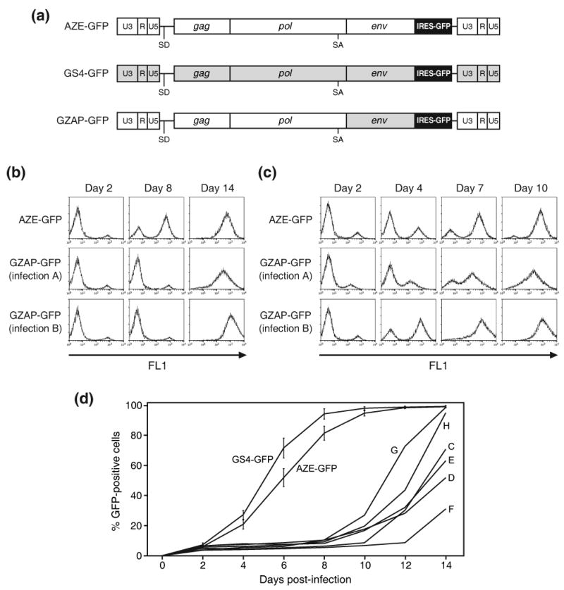
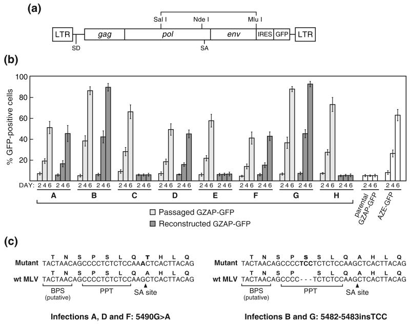
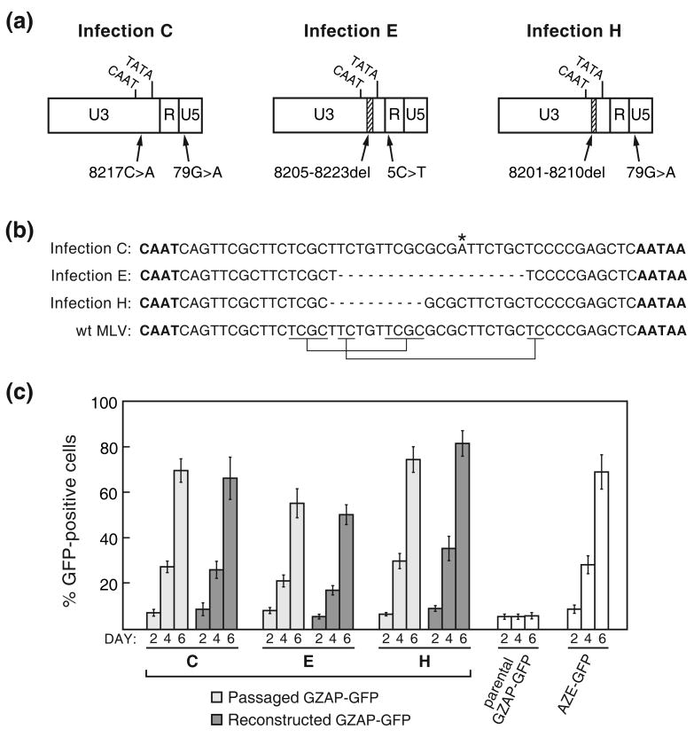
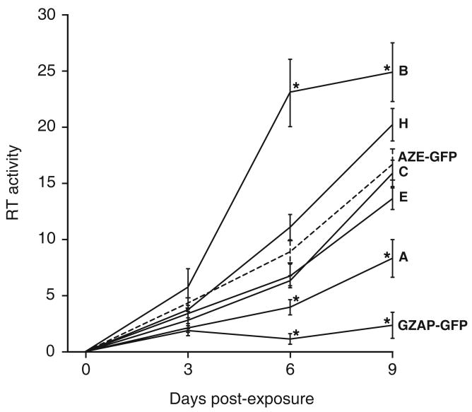
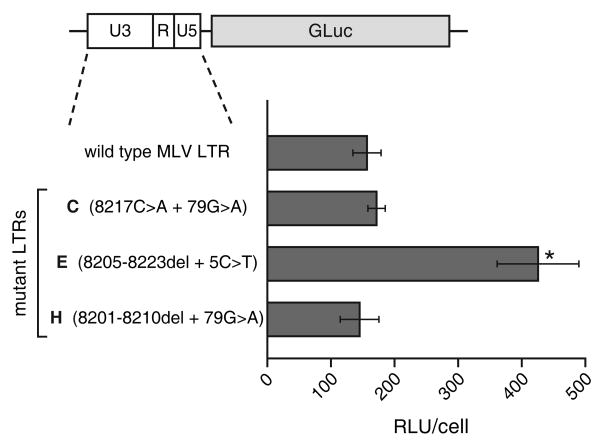
Retroviruses are well known for their ability to incorporate envelope (Env) proteins from other retroviral strains and genera, and even from other virus families. This characteristic has been widely exploited for the generation of replication-defective retroviral vectors, including those derived from murine leukemia virus (MLV), bearing heterologous Env proteins. We investigated the possibility of "genetically pseudotyping" replication-competent MLV by replacing the native env gene in a full-length viral genome with that of another gammaretrovirus. Earlier, we developed replication-competent versions of MLV that stably transmit and express transgenes inserted into the 3' untranslated region of the viral genome. In one such tagged MLV expressing green fluorescent protein, we replaced the native env sequence with that of gibbon ape leukemia virus (GALV). Although the GALV Env protein is commonly used to make high-titer pseudotypes of MLV vectors, we found that the env replacement greatly attenuated viral replication. However, extended cultivation of cells exposed to the chimeric virus resulted in selection of mutants exhibiting rapid replication kinetics and different variants arose in different infections. Two of these variants had acquired mutations at or adjacent to the splice acceptor site, and three others had acquired dual mutations within the long terminal repeat. Analysis of the levels of unspliced and spliced viral RNA produced by the parental and adapted viruses showed that the mutations gained by each of these variants functioned to reverse an imbalance in splicing caused by the env gene substitution. Our results reveal the presence of previously unknown cis-acting sequences in MLV that modulate splicing of the viral transcript and demonstrate that tagging of the retroviral genome with an easily assayed transgene can be combined with in vitro evolution as an approach to efficiently generating and screening for replicating mutants of replication-impaired recombinant viruses.


Figures

Figure 1
Figure 1
Structure and in vitro adaptation of an MLV-GALV chimera. (a) Schematic of the viruses used in this study. AZE-GFP and GS4-GFP are replication-competent clones of MLV and GALV, respectively, containing an IRES-GFP cassette between the env gene and 3′ UTR. GZAP-GFP is an MLV-GALV chimera identical to AZE-GFP except that it contains the env gene of GALV. Unshaded regions represent sequences derived from MLV and grey shaded regions represent those derived from GALV. (b) Histograms from flow cytometric analysis of primary infections with AZE-GFP or GZAP-GFP at equal MOI. LNCaP cells were exposed to supernatant from cells transfected with the provirus-containing plasmids and viral spread was monitored by GFP expression on the indicated days post-exposure. (c) Histograms from secondary infections of LNCaP cells carried out using supernatant from Day 14 of the infections shown in (b). (d) Results of additional primary infections of LNCaP cells with GZAP-GFP, AZE-GFP and GS4-GFP. The values for AZE-GFP and GS4-GFP are the averages obtained from three infections carried out in parallel and error bars represent standard deviations. Letters C through H denote single, independent infections with GZAP-GFP.
Figure 2
Figure 2
Screening of the eight passaged populations of GZAP-GFP for replication-enabling mutations in the 4.1-kb SalI-MluI pol-env region of the virus. (a) Schematic of the provirus showing the region that was amplified by PCR from genomic DNA and reintroduced into pGZAP-GFP for functional analysis. (b) Results from infections initiated with either passaged virus from the original eight infections or with virus produced by transfection of pGZAP-GFP reconstructed with the corresponding amplified fragments. LNCaP cells were exposed to an equal number of GFP-transducing units of virus from the original infections (“passaged GZAP-GFP”) or from transfections with reconstructed pGZAP-GFP, and spread was quantitated by flow cytometry at the indicated days post-infection. Infections with parental GZAP-GFP and AZE-GFP, produced by transfection, were included as controls. Values shown are the average obtained from triplicate infections and error bars indicate standard deviations. (c) The adaptive splice acceptor mutations that arose in GZAP-GFP during infections A, B, D, F and G. The mutant and wild type sequences around the acceptor site are shown aligned. Above each nucleotide sequence is the corresponding translation for integrase. The mutations and the amino acids that are altered are in bold. The locations of the MLV splice acceptor (SA) site, polypyrimidine tract (PPT), and putative branch point sequence (BPS) are indicated.
Figure 3
Figure 3
Replication-enabling mutations that arose in GZAP-GFP during infections C, E and H. (a) Schematic of the LTRs of the mutants showing the locations of deletions and point mutations identified by sequencing of proviral DNA. Hatched segments indicate deletions. (b) Alignment of the U3 region sequences between the CAAT and TATA boxes of each mutant with the corresponding wild type MLV sequence. The CAAT and TATA boxes are in bold, the 8217C>A mutation is marked by an asterisk, deleted nucleotides are represented by dashes and the short direct repeats that flanked the regions deleted in infections E and H are indicated below the wild type MLV sequence. (c) Comparison of the replication of virus from the passaged GZAP-GFP populations to that of GZAP-GFP reconstructed to contain the corresponding dual LTR mutations shown in (a). Values shown are the average obtained from triplicate infections and error bars indicate standard deviations.
Figure 4
Figure 4
Evaluation of the importance of each of the LTR mutations from infections C, E and H for viral replication. For each of these three GZAP-GFP mutants, two variants were constructed, each possessing one of the two mutations in isolation. Infections with these variants or the original mutants were carried out using equal doses of virus, and viral spread was assessed by flow cytometry at the indicated days post-infection. Values shown are the average obtained from triplicate infections and error bars indicate standard deviations.
Figure 5
Figure 5
Replication of viruses as measured by reverse transcriptase activity. LNCaP cells were exposed to each virus at an MOI of 0.1 with GZAP-GFP, the indicated GZAP-GFP variants, or AZE-GFP, and RT activity in the cultures was monitored over the following nine days. RT activities are expressed in arbitrary units and are the average obtained from triplicate infections. Error bars represent standard deviations. Asterisks denote RT activities that were significantly different (p < 0.05 by two-tailed, paired t-test) from those of AZE-GFP at the same time point. The values for AZE-GFP are shown by a dashed line for reference. The RT value for Day 0 represents the activity in cultures immediately before infection.
Figure 6
Figure 6
Comparison of the infectivity of the adapted chimeric viruses on human and mouse cells. PC-3 human prostate carcinoma and NIH3T3 mouse fibroblast cells were infected at a MOI of 0.1 and were analyzed by flow cytometry at 5 days post-infection. Vertical axis: cell number, horizontal axis: GFP fluorescence measured using the FL1 channel
Figure 7
Figure 7
Identification of the splice junction of GALV. RNA was isolated from cells transfected with either pGALV-S, which contains a wild type proviral clone of GALV, or pGS4-GFP and used for RT-PCR. A forward primer in U5 and a reverse primer immediately upstream of the env start codon were used to amplify across the splice junction. The amplified products were gel purified and directly sequenced. (a) Agarose gel of the RT-PCR reactions. (b) Alignment of the amplified sequences (top strand) with that of GALV genomic sequence (bottom strand). The GALV RNA genome coordinates at the 5′ and 3′ ends of the identified intron are shown. The GT dinucleotide of the splice donor and the AG of the splice acceptor are underlined.
Figure 8
Figure 8
Quantitation of the levels of spliced and unspliced viral RNA produced by MLV, GALV and chimeric viruses. RNA isolated from cells transfected with the provirus-containing plasmids was reverse transcribed and used in TaqMan PCR. (a) Schematic diagram of the gammaretroviral RNA genome showing locations of the primers (arrowheads) and probes (rectangles) used to measure the levels of unspliced and spliced transcript. The primer-probe sets above the RNA diagram represent the location of those used for wild type and GFP-tagged MLV and for GZAP-GFP and its mutants. The primer probe sets below the RNA diagram represent the location of those used for wild type and GFP-tagged GALV. Black circles indicate the locations of the splice donor and acceptor sites in the viral RNA. (b) Results of quantitation of the transcripts, expressed as the ratio of unspliced to spliced viral RNA. The ratio for wild type MLV (Moloney strain) was arbitrarily set to 1, and the ratios for all other viruses are normalized to this value. Each value is the mean ratio obtained from at least three amplifications, and error bars represent standard deviations. ZAPm-GFP is identical to AZE-GFP and GZAP-GFP but contains the ecotropic MLV env gene.
Figure 9
Figure 9
Comparison of the transcriptional activity of the wild type and mutant LTRs. LNCaP cells were transiently transfected with reporter plasmids containing the wild type or mutant LTRs linked to Gaussia luciferase. Shown for each construct are the average relative light units (RLU) produced per cell, after normalization for transfection efficiency. Values were obtained from four replicates and error bars represent standard deviations. The asterisk indicates a statistically significant difference between the activity of the LTR of mutant E and that of wild type virus (p = 0.012 by two-tailed, paired t-test).

Similar articles

Cited by

References

    1. Overbaugh J, Miller AD, Eiden MV. Receptors and entry cofactors for retroviruses include single and multiple transmembrane-spanning proteins as well as newly described glycophosphatidylinositol-anchored and secreted proteins. Microbiol Mol Biol Rev. 2001;65:371–89. - PMC - PubMed
    1. Sanders DA. No false start for novel pseudotyped vectors. Curr Opin Biotechnol. 2002;13:437–42. - PubMed
    1. Mild M, Esbjornsson J, Fenyo EM, Medstrand P. Frequent Intrapatient Recombination between Human Immunodeficiency Virus Type 1 R5 and X4 Envelopes: Implications for Coreceptor Switch. J Virol. 2007;81:3369–76. - PMC - PubMed
    1. Roy-Burman P. Endogenous env elements: partners in generation of pathogenic feline leukemia viruses. Virus Genes. 1995;11:147–61. - PubMed
    1. Chen BK, Rousso I, Shim S, Kim PS. Efficient assembly of an HIV-1/MLV Gag-chimeric virus in murine cells. Proc Natl Acad Sci U S A. 2001;98:15239–44. - PMC - PubMed

Publication types

MeSH terms

Substances