

A novel spectral tuning in the short wavelength-sensitive (SWS1 and SWS2) pigments of bluefin killifish (Lucania goodei)
- PMID: 17498892
- PMCID: PMC1963460
- DOI: 10.1016/j.gene.2007.03.019
A novel spectral tuning in the short wavelength-sensitive (SWS1 and SWS2) pigments of bluefin killifish (Lucania goodei)
Abstract
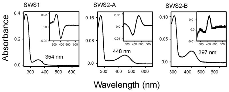
The molecular bases of spectral tuning in the UV-, violet-, and blue-sensitive pigments are not well understood. Using the in vitro assay, here we show that the SWS1, SWS2-A, and SWS2-B pigments of bluefin killifish (Lucania goodei) have the wavelengths of maximal absorption (lambda(max)'s) of 354, 448, and 397 nm, respectively. The spectral difference between the SWS2-A and SWS2-B pigments is largest among those of all currently known pairs of SWS2 pigments within a species. The SWS1 pigment contains no amino acid replacement at the currently known 25 critical sites and seems to have inherited its UV-sensitivity directly from the vertebrate ancestor. Mutagenesis analyses show that the amino acid differences at sites 44, 46, 94, 97, 109, 116, 118, 265, and 292 of the SWS2-A and SWS2-B pigments explain 80% of their spectral difference. Moreover, the larger the individual effects of amino acid changes on the lambda(max)-shift are, the larger the synergistic effects tend to be generated, revealing a novel mechanism of spectral tuning of visual pigments.
Figures

Similar articles
-
The spectral tuning in the short wavelength-sensitive type 2 pigments.Gene. 2003 Mar 13;306:91-8. doi: 10.1016/s0378-1119(03)00424-4. Gene. 2003. PMID: 12657470
-
Spectral tuning in vertebrate short wavelength-sensitive 1 (SWS1) visual pigments: can wavelength sensitivity be inferred from sequence data?J Exp Zool B Mol Dev Evol. 2014 Nov;322(7):529-39. doi: 10.1002/jez.b.22576. Epub 2014 Jun 2. J Exp Zool B Mol Dev Evol. 2014. PMID: 24890094 Review.
-
The molecular mechanism for the spectral shifts between vertebrate ultraviolet- and violet-sensitive cone visual pigments.Biochem J. 2002 Oct 1;367(Pt 1):129-35. doi: 10.1042/BJ20020483. Biochem J. 2002. PMID: 12099889 Free PMC article.
-
Molecular basis of spectral tuning in the newt short wavelength sensitive visual pigment.Biochemistry. 2003 May 27;42(20):6025-34. doi: 10.1021/bi020629+. Biochemistry. 2003. PMID: 12755604
-
Spectral tuning of shortwave-sensitive visual pigments in vertebrates.Photochem Photobiol. 2007 Mar-Apr;83(2):303-10. doi: 10.1562/2006-06-27-IR-952. Photochem Photobiol. 2007. PMID: 17576346 Review.
Cited by
-
Mechanisms of spectral tuning in the RH2 pigments of Tokay gecko and American chameleon.Gene. 2007 Sep 1;399(1):26-32. doi: 10.1016/j.gene.2007.04.036. Epub 2007 May 10. Gene. 2007. PMID: 17590287 Free PMC article.
-
Origin and adaptation of green-sensitive (RH2) pigments in vertebrates.FEBS Open Bio. 2020 May;10(5):873-882. doi: 10.1002/2211-5463.12843. Epub 2020 Apr 11. FEBS Open Bio. 2020. PMID: 32189477 Free PMC article.
-
Diurnal lighting patterns and habitat alter opsin expression and colour preferences in a killifish.Proc Biol Sci. 2013 May 22;280(1763):20130796. doi: 10.1098/rspb.2013.0796. Print 2013 Jul 22. Proc Biol Sci. 2013. PMID: 23698009 Free PMC article.
-
Orthologous Divergence and Paralogous Anticonvergence in Molecular Evolution of Triplicated Green Opsin Genes in Medaka Fish, Genus Oryzias.Genome Biol Evol. 2020 Jun 1;12(6):911-923. doi: 10.1093/gbe/evaa111. Genome Biol Evol. 2020. PMID: 32467976 Free PMC article.
-
Visual pigment evolution in Characiformes: The dynamic interplay of teleost whole-genome duplication, surviving opsins and spectral tuning.Mol Ecol. 2020 Jun;29(12):2234-2253. doi: 10.1111/mec.15474. Epub 2020 Jun 8. Mol Ecol. 2020. PMID: 32421918 Free PMC article.
References
-
- Asenjo AB, Rim J, Oprian DD. Molecular determination of human red/green color discrimination. Neuron. 1994;12:1131–1138. - PubMed
-
- Babu KR, Dukkipati A, Birge RR, Knox BE. Regulation of phototransduction in short-wavelength cone visual pigments via the retinylidene Schiff base counterion. Biochemistry. 2001;40:13760–13766. - PubMed
-
- Carroll SB. The Making of the Fittest. W. W. Norton; New York: 2006.
-
- Carvalho LDS, Cowing JA, Wilkie SE, Bowmaker JK, Hunt DM. Shortwave visual sensitivity in tree and flying squirrels reflects changes in lifestyle. Current Biol. 2006;16:R81–R83. - PubMed
-
- Chinen A, Matsumoto Y, Kawamura S. Reconstruction of ancestral green visual pigments of zebrafish and molecular mechanism of their spectral differentiation. Mol Biol Evol. 2005a;22:1001–1010. - PubMed
Publication types
MeSH terms
Substances
Grants and funding
LinkOut - more resources
Full Text Sources
Miscellaneous
