Review
. 2007;39(7-8):1476-82.
doi: 10.1016/j.biocel.2007.03.022. Epub 2007 Apr 8.

MDM2 and MDM4: p53 regulators as targets in anticancer therapy

Affiliations
Review

MDM2 and MDM4: p53 regulators as targets in anticancer therapy

Franck Toledo et al. Int J Biochem Cell Biol. 2007.

Abstract

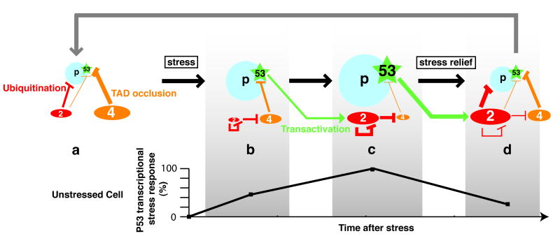
The gene TP53, encoding transcription factor p53, is mutated or deleted in half of human cancers, demonstrating the crucial role of p53 in tumor suppression. Importantly, p53 inactivation in cancers can also result from the amplification/overexpression of its specific inhibitors MDM2 and MDM4 (also known as MDMX). The presence of wild-type p53 in those tumors with MDM2 or MDM4 overexpression stimulates the search for new therapeutic agents to selectively reactivate it. This short survey highlights recent insights into MDM2 and MDM4 regulatory functions and their implications for the design of future p53-based anticancer strategies. We now know that MDM2 and MDM4 inhibit p53 in distinct and complementary ways: MDM4 regulates p53 activity, while MDM2 mainly regulates p53 stability. Upon DNA damage, MDM2-dependent degradation of itself and MDM4 contribute significantly to p53 stabilization and activation. These and other data imply that the combined use of MDM2 and MDM4 antagonists in cancer cells expressing wild-type p53 should activate p53 more significantly than agents that only antagonize MDM2, resulting in more effective anti-tumor activity.


Figures

Figure 1
Figure 1
Comparison of MDM2 and MDM4 primary structures. The p53-BoxI binding domain (BoxI BD; amino acids ca. 25-110), the Zinc finger domain (ZD; aa ca. 290-330) and the RING domain (RING; aa ca. 435-482) are conserved. The BoxI BD is the most conserved domain, and a sequence comparison of amino acids most important for interaction with p53 are shown, with residues that constitute the p53-binding hydrophobic pocket in bold (see text for details). A « lid » before the p53-BoxI BD (i; aa 16-24), which sequence is not conserved, is also proposed to regulate interactions with p53. Both proteins contain a region rich in acidic residues (Acid; aa 237-288 in MDM2, aa 215-255 in MDM4), but these regions do not share any significant sequence homology. The Acidic region in MDM2 is proposed to interact with the S9-S10 β sheets and BoxV from the p53 DNA binding domain, and is thus noted BV BD. L, nuclear localization signal; E, nuclear export signal.
Figure 2
Figure 2
A dynamic model of the p53 response integrating the distinct and complementary roles of MDM2 and MDM4. (a) in unstressed cells p53 is kept at low levels (due to MDM2-mediated degradation) and inactive (primarily due to MDM4-mediated occlusion of the p53 TAD). In this diagram, p53 stability is represented by a blue circle and p53 activity by a green star, and MDM2 (2) and MDM4 (4) levels are represented by red and orange ovals, respectively. (b) after stress, MDM2 degrades itself and MDM4: a transcriptional stress response (diagrammed below) is mounting. (c) increased MDM2 levels, resulting from p53 activation, lead to a more efficient MDM4 degradation, enabling full p53 activation: the transcriptional response is maximal. (d) following stress relief, accumulated MDM2 targets p53 again, and as MDM4 levels also increase, p53 activity decreases, so that the stress response is fading. This may allow cell cycle re-entry (grey arrow). As discussed in the text, the switch that makes MDM2 preferentially target p53 for degradation in unstressed cells (a), then target itself and MDM4 after stress (b and c) then target p53 again after stress relief (d) is proposed to result from the regulated deubiquitination of p53, MDM2 and MDM4 by HAUSP, a process also involving the adaptor protein Daxx. The model is adapted from (Toledo et al., 2006).

Similar articles

Cited by

References

    1. Bernal F, Tyler AF, Korsmeyer SJ, et al. Reactivation of the p53 Tumor Suppressor Pathway by a Stapled p53 Peptide. J Am Chem Soc. 2007;129:2456–2457. - PMC - PubMed
    1. Boesten LS, Zadelaar SM, De Clercq S, et al. Mdm2, but not Mdm4, protects terminally differentiated smooth muscle cells from p53-mediated caspase-3-independent cell death. Cell Death Differ. 2006;13:2089–2098. - PubMed
    1. Brummelkamp TR, Fabius AW, Mullenders J, et al. An shRNA barcode screen provides insight into cancer cell vulnerability to MDM2 inhibitors. Nat Chem Biol. 2006;2:202–206. - PubMed
    1. Chene P. Inhibition of the p53-MDM2 interaction: targeting a protein-protein interface. Mol Cancer Res. 2004;2:20–28. - PubMed
    1. Chipuk JE, Green DR. Dissecting p53-dependent apoptosis. Cell Death Differ. 2006;13:994–1002. - PubMed

Publication types

MeSH terms