

The role of the pre-supplementary motor area in the control of action
- PMID: 17499162
- PMCID: PMC2648723
- DOI: 10.1016/j.neuroimage.2007.03.034
The role of the pre-supplementary motor area in the control of action
Abstract
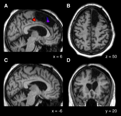
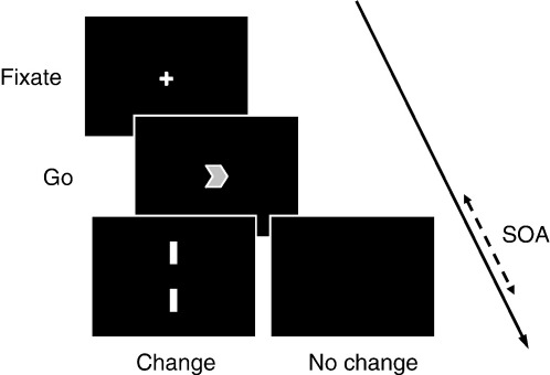
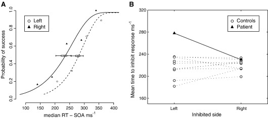
Although regions within the medial frontal cortex are known to be active during voluntary movements their precise role remains unclear. Here we combine functional imaging localisation with psychophysics to demonstrate a strikingly selective contralesional impairment in the ability to inhibit ongoing movement plans in a patient with a rare lesion involving the right pre-supplementary motor area (pre-SMA), but sparing the supplementary motor area (SMA). We find no corresponding delay in simple reaction times, and show that the inhibitory deficit is sensitive to the presence of competition between responses. The findings demonstrate that the pre-SMA plays a critical role in exerting control over voluntary actions in situations of response conflict. We discuss these findings in the context of a unified framework of pre-SMA function, and explore the degree to which extant data on this region can be explained by this function alone.
Figures
Similar articles
-
Human medial frontal cortex mediates unconscious inhibition of voluntary action.Neuron. 2007 Jun 7;54(5):697-711. doi: 10.1016/j.neuron.2007.05.016. Neuron. 2007. PMID: 17553420 Free PMC article.
-
Cortical connectivity after subcortical stroke assessed with functional magnetic resonance imaging.Ann Neurol. 2008 Feb;63(2):236-46. doi: 10.1002/ana.21228. Ann Neurol. 2008. PMID: 17896791
-
Supplementary motor area activation in patients with frontal lobe tumors and arteriovenous malformations.AJNR Am J Neuroradiol. 2003 Oct;24(9):1837-42. AJNR Am J Neuroradiol. 2003. PMID: 14561613 Free PMC article.
-
Activation likelihood estimation meta-analysis of motor-related neural activity after stroke.Neuroimage. 2012 Feb 1;59(3):2771-82. doi: 10.1016/j.neuroimage.2011.10.023. Epub 2011 Oct 17. Neuroimage. 2012. PMID: 22023742 Review.
-
[Surgical treatments for the patients with SMA tumor].No Shinkei Geka. 2005 Apr;33(4):327-36. No Shinkei Geka. 2005. PMID: 15830538 Review. Japanese. No abstract available.
Cited by
-
Dynamical EEG Indices of Progressive Motor Inhibition and Error-Monitoring.Brain Sci. 2021 Apr 9;11(4):478. doi: 10.3390/brainsci11040478. Brain Sci. 2021. PMID: 33918711 Free PMC article.
-
Functional magnetic resonance imaging multivoxel pattern analysis reveals neuronal substrates for collaboration and competition with myopic and predictive strategic reasoning.Hum Brain Mapp. 2020 Oct 15;41(15):4314-4331. doi: 10.1002/hbm.25127. Epub 2020 Jul 7. Hum Brain Mapp. 2020. PMID: 32633451 Free PMC article.
-
Functional anatomy of idiomatic expressions.Brain Topogr. 2021 Jul;34(4):489-503. doi: 10.1007/s10548-021-00843-3. Epub 2021 May 4. Brain Topogr. 2021. PMID: 33948754
-
Are metaphors embodied? The neural evidence.Psychol Res. 2022 Nov;86(8):2417-2433. doi: 10.1007/s00426-021-01604-4. Psychol Res. 2022. PMID: 34762153 Free PMC article.
-
Altered intrahemispheric structural connectivity in Gilles de la Tourette syndrome.Neuroimage Clin. 2013 Dec 7;4:174-81. doi: 10.1016/j.nicl.2013.11.011. eCollection 2014. Neuroimage Clin. 2013. PMID: 24371800 Free PMC article.
References
-
- Alexander G.E., Crutcher M.D. Preparation for movement – neural representations of intended direction in 3 motor areas of the monkey. J. Neurophysiol. 1990;64:133–150. - PubMed
-
- Amador N., Fried I. Single-neuron activity in the human supplementary motor area underlying preparation for action. J. Neurosurg. 2004;100:250–259. - PubMed
-
- Baird A., Dewar B.K., Critchley H., Gilbert S.J., Dolan R.J., Cipolotti L. Cognitive functioning after medial frontal lobe damage including the anterior cingulate cortex: a preliminary investigation. Brain Cogn. 2006;60:166–175. - PubMed
-
- Behrens T.E., Jenkinson M., Robson M.D., Smith S.M., Johansen-Berg H. A consistent relationship between local white matter architecture and functional specialisation in medial frontal cortex. NeuroImage. 2006;30:220–227. - PubMed
Publication types
MeSH terms
Grants and funding
LinkOut - more resources
Full Text Sources
Medical
