Spontaneous myocarditis mimicking human disease occurs in the presence of an appropriate MHC and non-MHC background in transgenic mice
- PMID: 17499268
- PMCID: PMC1993806
- DOI: 10.1016/j.yjmcc.2007.03.898
Spontaneous myocarditis mimicking human disease occurs in the presence of an appropriate MHC and non-MHC background in transgenic mice
Abstract
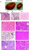
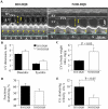
Most individuals have viral infections at some point in their life, however, only few develop autoreactivity to cardiac myosin following infection resulting in myocarditis suggesting a genetic predisposition. Most mouse models of myocarditis are induced by viral infection or by immunization with cardiac myosin. We generated HLA-DR3.Abetao and HLA-DQ8.Abetao transgenic mice in NOD and HLA-DQ8.Abetao in B10 background to study spontaneous autoimmunity. A high mortality was observed in NOD.DQ8 female mice 16 weeks or older. Echocardiography showed marked systolic dysfunction. Histopathology of various organs revealed an enlarged heart with mononuclear infiltrate consisting of CD4 and Mac-1+ cells and myocyte necrosis. The autoimmunity was associated with the presence of spontaneous autoreactive T cells and antibodies to cardiac myosin. Serologically, mice were negative for all known mouse viruses. NOD.DR3.Abetao, the transgene negative littermates, NOD, and B10.DQ8 Abetao mice had no gross or microscopic cardiac pathology. Spontaneous cellular and humoral response to cardiac myosin suggests that NOD.DQ8 may harbor autoreactive cells that can lead to spontaneous myocarditis and dilated cardiomyopathy. HLA-DQ8 is required for the predisposition to the spontaneous autoreactivity while NOD background influences onset and progression of disease. This model of myocarditis occurs predominantly in female mice and may provide insight into the pathogenesis of heart disease in women.
Figures


Similar articles
-
Spontaneous autoimmune myocarditis and cardiomyopathy in HLA-DQ8.NODAbo transgenic mice.J Autoimmun. 2009 Nov-Dec;33(3-4):260-9. doi: 10.1016/j.jaut.2009.09.005. Epub 2009 Oct 7. J Autoimmun. 2009. PMID: 19811893 Free PMC article.
-
CD4 T cells play major effector role and CD8 T cells initiating role in spontaneous autoimmune myocarditis of HLA-DQ8 transgenic IAb knockout nonobese diabetic mice.J Immunol. 2006 Jun 15;176(12):7715-25. doi: 10.4049/jimmunol.176.12.7715. J Immunol. 2006. PMID: 16751419
-
A spontaneous model for autoimmune myocarditis using the human MHC molecule HLA-DQ8.J Immunol. 2004 Feb 15;172(4):2651-8. doi: 10.4049/jimmunol.172.4.2651. J Immunol. 2004. PMID: 14764740
-
Pathogenesis of myocarditis and dilated cardiomyopathy.Adv Immunol. 2008;99:95-114. doi: 10.1016/S0065-2776(08)00604-4. Adv Immunol. 2008. PMID: 19117533 Review.
-
T cell mimicry in inflammatory heart disease.Mol Immunol. 2004 Feb;40(14-15):1121-7. doi: 10.1016/j.molimm.2003.11.023. Mol Immunol. 2004. PMID: 15036918 Review.
Cited by
-
FTY720 (Gilenya) treatment prevents spontaneous autoimmune myocarditis and dilated cardiomyopathy in transgenic HLA-DQ8-BALB/c mice.Cardiovasc Pathol. 2016 Sep-Oct;25(5):353-61. doi: 10.1016/j.carpath.2016.05.003. Epub 2016 May 17. Cardiovasc Pathol. 2016. PMID: 27288745 Free PMC article.
-
Impaired thymic tolerance to α-myosin directs autoimmunity to the heart in mice and humans.J Clin Invest. 2011 Apr;121(4):1561-73. doi: 10.1172/JCI44583. Epub 2011 Mar 23. J Clin Invest. 2011. PMID: 21436590 Free PMC article.
-
Spontaneous autoimmune myocarditis and cardiomyopathy in HLA-DQ8.NODAbo transgenic mice.J Autoimmun. 2009 Nov-Dec;33(3-4):260-9. doi: 10.1016/j.jaut.2009.09.005. Epub 2009 Oct 7. J Autoimmun. 2009. PMID: 19811893 Free PMC article.
-
[Etiology, diagnosis, management, and treatment of myocarditis. Position paper from the ESC Working Group on Myocardial and Pericardial Diseases].Herz. 2013 Dec;38(8):855-61. doi: 10.1007/s00059-013-3988-7. Herz. 2013. PMID: 24165990 German.
-
Myocarditis in Humans and in Experimental Animal Models.Front Cardiovasc Med. 2019 May 16;6:64. doi: 10.3389/fcvm.2019.00064. eCollection 2019. Front Cardiovasc Med. 2019. PMID: 31157241 Free PMC article. Review.
References
-
-
Kim KS, Hofling K, Carson SD, Chapman NM, Tracy S. The primary viruses of Myocarditis. Humana Press; NJ: 2002. pp. PP23–53. In edited book: Myocarditis.
-
-
- Olinde KD, O’ Connel JB. Inflammatory heart disease: pathogenesis, clinical manifestations, and treatment of myocarditis. Annu. Rev. Med. 1994;45:481–90. - PubMed
-
- Maisch B, Bauer E, Cirsi M, Kochsiek K. Cytolytic cross-reactive antibodies directed against the cardiac membrane and viral proteins in coxsackievirus B3 and B4 myocarditis. Characterization and pathogenetic relevance. Circulation. 1993;87:49–65. - PubMed
-
- Caforio AL, Goldman JH, Haven AJ, Baig KM, McKenna WJ. Evidence for autoimmunity to myosin and other heart specific autoantigens in patients with dilated cardiomyopathy. Int. J. Cardiol. 1996;54:157–63. - PubMed
-
- Limas CJ. Autoimmunity in dilated cardiomyopathy and the major histocompatibility complex. Int. J. Cardiol. 1996;54:113–6. - PubMed
Publication types
MeSH terms
Substances
Grants and funding
LinkOut - more resources
Full Text Sources
Molecular Biology Databases
Research Materials

