. 2007 Jun 29;370(1):157-69.
doi: 10.1016/j.jmb.2007.04.049. Epub 2007 May 4.

Identification of a single HNH active site in type IIS restriction endonuclease Eco31I

Affiliations

Identification of a single HNH active site in type IIS restriction endonuclease Eco31I

Arturas Jakubauskas et al. J Mol Biol. .

Abstract

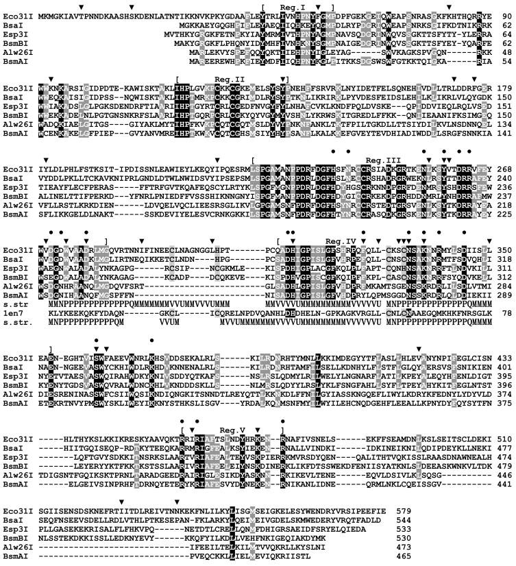
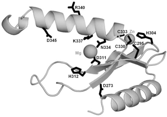
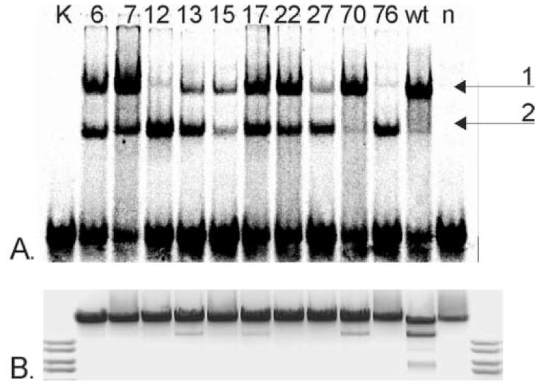
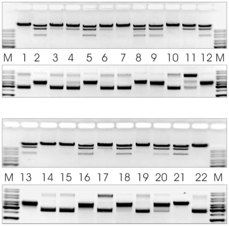
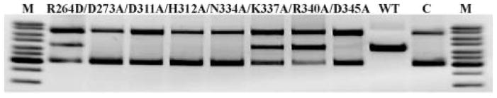
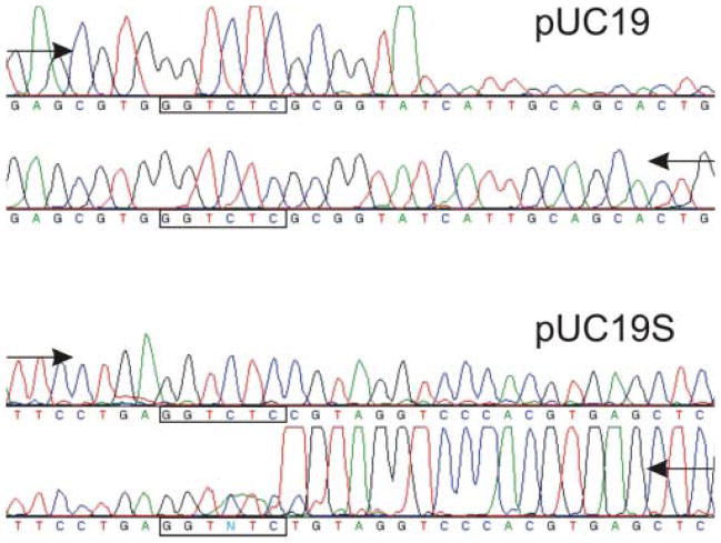
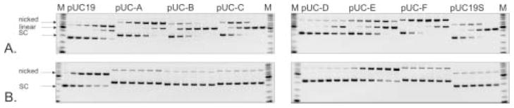
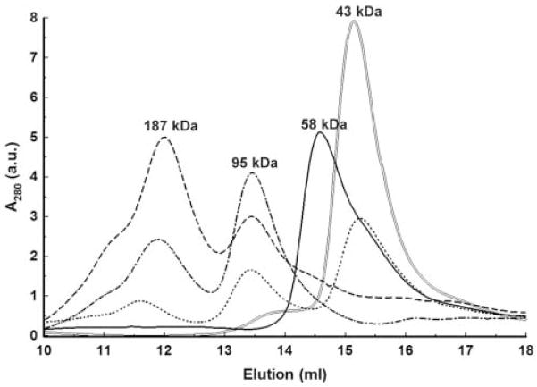
Type IIS restriction endonuclease Eco31I is a "short-distance cutter", which cleaves DNA strands close to its recognition sequence, 5'-GGTCTC(1/5). Previously, it has been proposed that related endonucleases recognizing a common sequence core GTCTC possess two active sites for cleavage of both strands in the DNA substrate. Here, we present bioinformatic identification and experimental evidence for a single nuclease active site. We identified a short region of homology between Eco31I and HNH nucleases, constructed a three-dimensional model of the putative catalytic domain and validated our predictions by random and site-specific mutagenesis. The restriction mechanism of Eco31I is suggested by analogy to the mechanisms of phage T4 endonuclease VII and homing endonuclease I-PpoI. We propose that residues D311 and N334 coordinate the cofactor. H312 acts as a general base-activating water molecule for the nucleophilic attack. K337 together with R340 and D345 are located in close proximity to the active center and are essential for correct folding of catalytic motif, while D345 together with R264 and D273 could be directly involved in DNA binding. We also predict that the Eco31I catalytic domain contains a putative Zn-binding site, which is essential for its structural integrity. Our results suggest that the HNH-like active site is involved in the cleavage of both strands in the DNA substrate. On the other hand, analysis of site-specific mutants in the region, previously suggested to harbor the second active site, revealed its irrelevance to the nuclease activity. Thus, our data argue against the earlier prediction and indicate the presence of a single conserved active site in type IIS restriction endonucleases that recognize common sequence core GTCTC.


Figures

Figure 1
Figure 1. Alignment of Eco31I and related REases recognizing a common pentanucleotide GTCTC
Residues conserved in 100% and >50% sequences are indicated by white color with black and grey shading, respectively. Mutations obtained by random mutagenesis are indicated by ‘▼;’; mutations introduced by site-directed mutagenesis are indicated by ‘●’. The middle panel indicates the predicted ‘His-Me finger’ structure with the HNH-like motif, aligned to the T4 Endonuclease VII structure (1en7). Secondary structures observed in 1en7 and predicted for Eco31I are shown (helices as tubes, strands as arrows). Additionally, the common functionally important residues are shaded in T4 Endonuclease VII sequence. Residues participating in the Zn-binding site (observed in 1en7, predicted for REases) are indicated by light grey shading.
Figure 2
Figure 2. Model of the ‘His-Me finger’ structure and the HNH-like active site of Eco31I (residues 271-354)
The protein backbone is shown as a grey ribbon. Side-chains of selected functionally important residues are shown in the wireframe representation and labeled. The predicted positions of Mg2+ and Zn2+ ions are indicated by balls.
Figure 3
Figure 3. Functional characteristics of primary Eco31I mutants. (A.) DNA-binding, (B.) DNA restriction activity (invert image)
Mutant numbers are indicated above the lanes. K, control DNA; wt, cell extract with wt Eco31I; n, cell extract without enzyme.
Figure 4
Figure 4. Restriction activity of secondary Eco31I mutants on λ DNA and pBR322 (invert images)
M, DNA molecular weight marker. Lane 1, m6-L303S/S332P/K337E; lane 2, m7-W274C; lane 3, m7-S361P; lane 4, m12-F326S/L350F; lane 5, m12-K472E; lane 6, m13-C333S; lane 7, m15-C333R; lane 8, m15-I459V; lane 9, m17-K82R/V270M; lane 10, m17-V270M/N334D; lane 11, m17-N334D; lane 12, m27-Y181F; lane 13, m27-Y181F/V260D; lane 14, m27-V260D/L329P; lane 15, m27-L329P; lane 16, m70-Y138F; lane 17, m70-L256P; lane 18, m76-I214T/Y259C; lane 19, m76-Y259C/D278G/I289V/F363L; lane 20, m76-D278G/I289V/F363L; lane 21, wt Eco31I; lane 22, cell extract without enzyme.
Figure 5
Figure 5. Restriction activity of site-directed Eco31I variants on pBR322 (invert image)
M, DNA molecular weight marker; WT, cell extract with wt Eco31I; C, cell extract without enzyme. Mutants are indicated above the lines.
Figure 6
Figure 6. Run-off sequencing to determine the nicking activity of Eco31I-N334D
The drop in peak signal indicates where the DNA polymerase runs off the template at the nicked site. Arrows indicate direction of DNA synthesis. Modified DNA polymerase adds an additional adenine (A) at the end of the extension product.
Figure 7
Figure 7. Reaction courses of Eco31I variants on plasmid DNA substrates (invert images)
M, DNA molecular weight marker; c(enzyme) = 100 μM; c(pDNA) = 20 nM. First lane of each reaction indicates zero point. Before loading the samples were pre-heated at 75°C for 20 min to avoid shifted zones. A. Reaction course of Eco31I-N334D. Aliquots were withdrawn at 2, 5, 10, 30 and 60 min. B. Reaction course of Eco31I-D311A. Aliquots were withdrawn at 1, 2, 3, 4 and 5 h.
Figure 8
Figure 8. Gel-filtration of Eco31I and its complexes with DNA
The numbers above the peaks denote the apparent molecular mass values, calculated by interpolating measured elution volumes onto the calibration curve. a.u. – arbitrary units. ‘—’ elution of free Eco31I; ‘=’ elution of free DNA; ‘■ ■ ■ ■’ elution of Eco31I-DNA (0.25–0.5 μM) complex; ‘- ■ -’ elution of Eco31I-DNA (1.0–0.5 μM) complex; ‘- - -’ elution of Eco31I-DNA (2.0–0.5 μM) complex.

Similar articles

Cited by

References

    1. Roberts RJ, Belfort M, Bestor T, Bhagwat AS, Bickle TA, Bitinaite J, Blumenthal RM, Degtyarev S, Dryden DT, Dybvig K, Firman K, Gromova ES, Gumport RI, Halford SE, Hattman S, Heitman J, Hornby DP, Janulaitis A, Jeltsch A, Josephsen J, Kiss A, Klaenhammer TR, Kobayashi I, Kong H, Kruger DH, Lacks S, Marinus MG, Miyahara M, Morgan RD, Murray NE, Nagaraja V, Piekarowicz A, Pingoud A, Raleigh E, Rao DN, Reich N, Repin VE, Selker EU, Shaw PC, Stein DC, Stoddard BL, Szybalski W, Trautner TA, Van Etten JL, Vitor JM, Wilson GG, Xu SY. A nomenclature for restriction enzymes, DNA methyltransferases, homing endonucleases and their genes. Nucleic Acids Res. 2003;31:1805–12. - PMC - PubMed
    1. Li L, Wu LP, Chandrasegaran S. Functional domains in Fok I restriction endonuclease. Proc Natl Acad Sci U S A. 1992;89:4275–9. - PMC - PubMed
    1. Wah DA, Hirsch JA, Dorner LF, Schildkraut I, Aggarwal AK. Structure of the multimodular endonuclease FokI bound to DNA. Nature. 1997;388:97–100. - PubMed
    1. Bitinaite J, Wah DA, Aggarwal AK, Schildkraut I. FokI dimerization is required for DNA cleavage. Proc Natl Acad Sci U S A. 1998;95:10570–5. - PMC - PubMed
    1. Zaremba M, Urbanke C, Halford SE, Siksnys V. Generation of the BfiI restriction endonuclease from the fusion of a DNA recognition domain to a non-specific nuclease from the phospholipase D superfamily. J Mol Biol. 2004;336:81–92. - PubMed

Publication types

MeSH terms

Substances

LinkOut - more resources