. 2007 Sep 15;366(1):62-72.
doi: 10.1016/j.virol.2007.03.060. Epub 2007 May 17.

Vaccinia virus temperature-sensitive mutants in the A28 gene produce non-infectious virions that bind to cells but are defective in entry

Affiliations

Vaccinia virus temperature-sensitive mutants in the A28 gene produce non-infectious virions that bind to cells but are defective in entry

Peter C Turner et al. Virology. .

Abstract

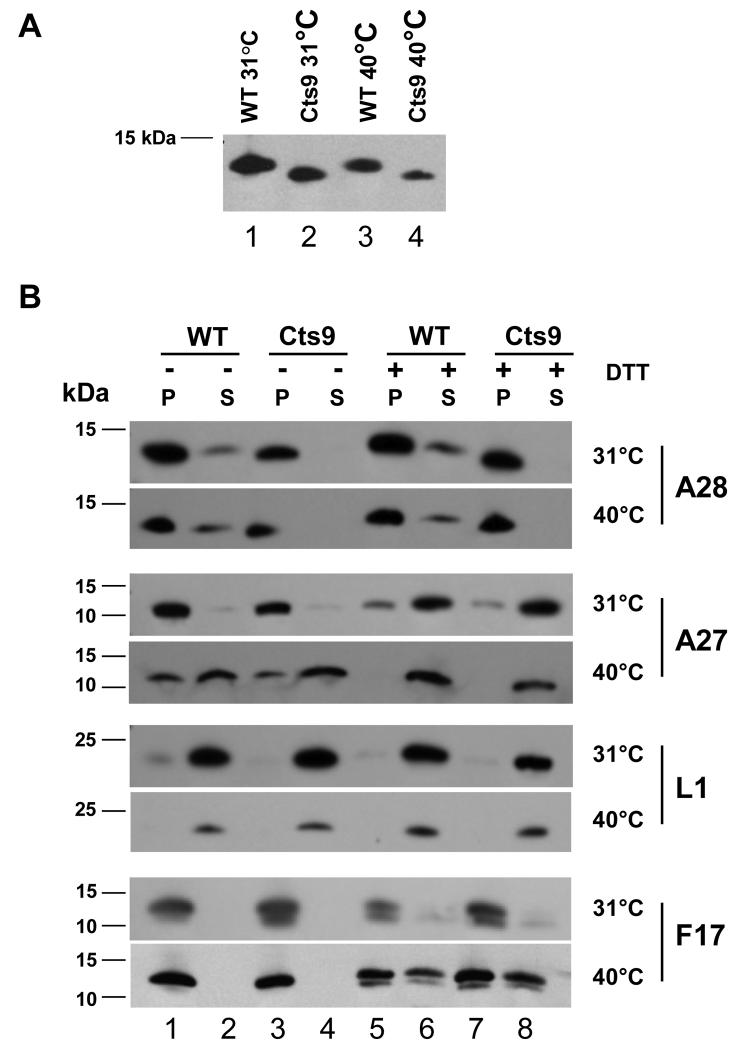
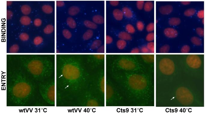
The vaccinia virus temperature-sensitive mutations Cts6 and Cts9 were mapped by marker rescue and DNA sequencing to the A28 gene. Cts6 and Cts9 contain an identical 2-bp deletion truncating the A28 protein and removing the fourth conserved cysteine near the C-terminus. Cts9 mutant virions produced at 40 degrees C were non-infectious and unable to cause cytopathic effect. However, the mutant A28 protein localized to purified mature virions (MV) at 31 degrees C and 40 degrees C. MV of Cts9 produced at 40 degrees C bound to cells but did not enter cells. Low pH treatment of Cts9-infected cells at 18 h p.i. failed to produce fusion from within at 40 degrees C, but gave fusion at 31 degrees C. Adsorption of Cts9 mutant virions to cells followed by low pH treatment showed a defect in fusion from without. The Cts9 phenotype suggests that the A28 protein is involved in both virus entry and cell-cell fusion, and supports the linkage between the two processes.


Figures

Figure 1
Figure 1. Marker rescue of Cts6
At the top is a diagram of a 7 kb region of the vaccinia genome within which Cts6 has been previously mapped, including genes A25L through A29L. Genes are represented by arrows, with the gene name and protein function indicated above. The horizontal lines immediately below represent a subset of PCR products used in the rescue of Cts6. The gene(s) included in each PCR product are indicated to the right of the product; LM32 corresponds to a 5 kb PCR product used in preliminary studies. The bottom of the figure shows selected crystal violet stained 60-mm dishes resulting from the marker rescue, with the corresponding PCR product labeled below the dish. Note that within the region comprising the non-essential genes A25L and A26L, the Copenhagen DNA sequence, from which the familiar gene nomenclature is derived, differs significantly from the WR sequence. In WR, the A type inclusion (ATI) gene is fragmented into 4 ORFs which we have called A25L, A25.1L, A25.2L and A25.3L. These 4 ORFs correspond to VACWR145, VACWR146, VACWR147 and VACWR148 respectively in the Poxvirus Bioinformatics Resource Center database (www.poxvirus.org). In the Copenhagen sequence, the A26L ORF is formed by a large deletion which spans A25.2 and A25.3 and fuses the N-terminus of the p4c gene with sequence near the 3' end of the ATI gene corresponding to the 3' end of the A25.1L ORF. The A25L ORF in WR and Copenhagen are approximately equivalent.
Figure 2
Figure 2. Sequences of the A28 gene and protein from wild type VV and from Cts6 and Cts9
A) Nucleotide alignment of the 3' end of the A28L gene from wild type, Cts6, and Cts9 viruses. In Cts6 and Cts9 cytosine residues located at positions 394 and 395 are deleted, which are indicated by ^ ^ below the sequence. B) Amino acid sequence alignment of A28 protein from wild type, Cts6, and Cts9. The first 131 amino acids are identical in mutant and wild type viruses. The deletion of the two adjacent C residues causes a frameshift which results in three amino acid substitutions (indicated by * under the sequence) followed by a premature stop codon causing truncation. Conserved cysteine residues are indicated by “C” in boldface. The A28 protein in our laboratory isolate of vaccinia WR was found to contain asparagine at position 124 rather than the aspartic acid expected from the published sequence (Senkevich et al., 2004a). A similar N124 substitution relative to the published VV WR sequence was found in the A28 protein of VV Copenhagen (Goebel et al., 1990) and of VV strain MVA. C) A diagram of the wt and mutant A28 proteins. A transmembrane domain, indicated by a gray box and labeled TM, is located between the 4th and 26th amino acids. The open box at the C-terminal end of the mutant protein represents the three substitutions encoded by the mutant viruses, which begin at amino acid 132. The Cs inside the proteins represent conserved cysteines.
Figure 3
Figure 3. Immunoblot analysis of purified wild type and mutant virions
A. Different mobilities of A28 protein from WT and Cts9 virions. Virions were incubated in the presence of 50 mM Tris for 30 minutes at 37°C, pelleted in a microfuge for 30 minutes, and subjected to electrophoresis on a 10-20% acrylamide Tris-tricine gel before immunoblotting with anti-A28 antibody. The nature of the A28 allele (WT or Cts9) and the temperature at which the virions were produced are shown above each lane. B. Western blot analysis of protein distribution in fractionated virions. Wild type (WT) or mutant (Cts9) virions grown at 31°C or 40°C were separated in the absence (−) or presence (+) of DTT into core (pellet, P) and membrane (supernatant, S) fractions and run on a 10-20% Tris-Tricine gel. Proteins were subjected to Western blot analysis with anti-A28, anti-A27, anti-L1, or anti-F17 antibody, as indicated on the right. Molecular weight markers are indicated on the left.
Figure 4
Figure 4. Lack of cytoplasmic effect following infection with Cts9 virions produced at the non-permissive temperature
BSC-40 cells were mock-infected (A), or infected at 31°C with purified MV of wtVV grown at 40°C (B), with Cts9 virions grown at 31°C (C), or with Cts9 virions produced at 40°C (D). At 6h p.i., the cells were fixed and stained for actin with phalloidin-AlexFluor 568.
Figure 5
Figure 5. Binding of Cts9 virions to cells but lack of entry
The upper four panels show binding to CV-1 cells of purified mature virions of either WT VV or Cts9 grown at 31°C or 40°C. CV-1 cells were infected at a multiplicity of 10 (for WT VV or Cts9 grown at 31°C), or for Cts9 grown at 40°C with an equivalent number of virus particles. Following adsorption of virions to cells for 1h at room temperature, the cells were fixed and reacted with anti-A27 mAb followed by goat anti-mouse FITC secondary Ab. The virions were pseudocolored cyan. Nuclei were counterstained by propidium iodide (red). The lower four panels show the result of the virus entry assay. Mature virions were adsorbed to cells at RT, and the cells transferred to 31°C for 2h to allow virus entry to occur. The cells were fixed, permeabilized, and stained with rabbit anti-A4 serum followed by goat anti-rabbit FITC (green) to visualize intracellular cores. Counterstaining of nuclei was with propidium iodide. The cells were photographed with a Zeiss Axiovert 200M inverted fluorescence microscope. Arrows in the lower panels indicate representative core particles.
Figure 6
Figure 6. Fusion from within by Cts9, and effect of temperature and multiplicity
A. Confluent BSC-40 cells were infected at moi = 5 with either wtVV (first two columns) or Cts9 (last two columns) at 31°C (top row) or 40°C (lower row). At 16h p.i., the monolayers were treated for 2 min with PBS at pH 7.4 or pH 4.6, as indicated above the panels, and incubated for a further 3h in complete medium before phase contrast photomicrography. B. Effect of multiplicity on fusion mediated by wtVV or Cts9 at 31°C. BSC-40 cells were infected with wtVV (upper row) or Cts9 (lower row) at a multiplicity of 50 (left column), 5 (center column), or 0.5 (right column), incubated for 16h at 31°C, briefly acid-treated at pH 4.6, and photographed after 3h incubation in medium.
Figure 7
Figure 7. Fusion from without
BSC-40 monolayers were mock-infected (A), or infected at moi = 300 (or with an equivalent number of non-infectious particles) in the presence of cycloheximide with purified MV of wtVV grown at 31°C (B, C) or with wtVV grown at 40°C (D), or with Cts9 produced at 31°C (E) or 40°C (F). Following virus adsorption and removal, all monolayers were briefly treated at pH 4.6 except for panel B, where monolayers were treated at pH 7.4. The cells were stained at 3h post-treatment with DAPI for nuclei (blue) and with phalloidin-AlexaFluor 568 for actin (red).

References

    1. Ausubel FM, Brent R, Kingston RE, Moore DD, Seidman JG, Smith JA, Struhl K. Current Protocols in Molecular Biology. John Wiley and Sons, Inc.; New York: 1994.
    1. Brown E, Senkevich TG, Moss B. Vaccinia virus F9 virion membrane protein is required for entry but not virus assembly, in contrast to the related L1 protein. J Virol. 2006;80:9455–9464. - PMC - PubMed
    1. Carter GC, Law M, Hollinshead M, Smith GL. Entry of the vaccinia virus intracellular mature virion and its interactions with glycosaminoglycans. J Gen.Virol. 2005;86:1279–1290. - PubMed
    1. Chung CS, Hsiao JC, Chang YS, Chang W. A27L protein mediates vaccinia virus interaction with cell surface heparan sulfate. J Virol. 1998;72:1577–1585. - PMC - PubMed
    1. Condit RC, Motyczka A. Isolation and preliminary characterization of temperature-sensitive mutants of vaccinia virus. Virology. 1981;113:224–241. - PubMed

Publication types

MeSH terms