

Matrices and scaffolds for drug delivery in dental, oral and craniofacial tissue engineering
- PMID: 17499385
- PMCID: PMC4035021
- DOI: 10.1016/j.addr.2007.03.019
Matrices and scaffolds for drug delivery in dental, oral and craniofacial tissue engineering
Abstract
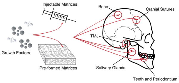
Current treatments for diseases and trauma of dental, oral and craniofacial (DOC) structures rely on durable materials such as amalgam and synthetic materials, or autologous tissue grafts. A paradigm shift has taken place to utilize tissue engineering and drug delivery approaches towards the regeneration of these structures. Several prototypes of DOC structures have been regenerated such as temporomandibular joint (TMJ) condyle, cranial sutures, tooth structures and periodontium components. However, many challenges remain when taking in consideration the high demand for esthetics of DOC structures, the complex environment and yet minimal scar formation in the oral cavity, and the need for accommodating multiple tissue phenotypes. This review highlights recent advances in the regeneration of DOC structures, including the tooth, periodontium, TMJ, cranial sutures and implant dentistry, with specific emphasis on controlled release of signaling cues for stem cells, biomaterial matrices and scaffolds, and integrated tissue engineering approaches.
Figures
Similar articles
-
Tissue engineering in dentistry.J Dent. 2014 Aug;42(8):915-28. doi: 10.1016/j.jdent.2014.05.008. Epub 2014 May 28. J Dent. 2014. PMID: 24880036 Review.
-
The crowning achievement: getting to the root of the problem.J Dent Educ. 2005 May;69(5):555-70. J Dent Educ. 2005. PMID: 15897336 Review.
-
Craniofacial tissue engineering by stem cells.J Dent Res. 2006 Nov;85(11):966-79. doi: 10.1177/154405910608501101. J Dent Res. 2006. PMID: 17062735 Free PMC article. Review.
-
Status and potential commercial impact of stem cell-based treatments on dental and craniofacial regeneration.Stem Cells Dev. 2006 Dec;15(6):881-7. doi: 10.1089/scd.2006.15.881. Stem Cells Dev. 2006. PMID: 17253950 Review.
-
Approaches to neural tissue engineering using scaffolds for drug delivery.Adv Drug Deliv Rev. 2007 May 30;59(4-5):325-38. doi: 10.1016/j.addr.2007.03.014. Epub 2007 Apr 10. Adv Drug Deliv Rev. 2007. PMID: 17482308 Free PMC article. Review.
Cited by
-
Comparison of TGFbR2 down-regulation in expanded HSCs on MBA/DBM scaffolds coated by UCB stromal cells.In Vitro Cell Dev Biol Anim. 2015 May;51(5):495-506. doi: 10.1007/s11626-014-9854-y. Epub 2014 Dec 25. In Vitro Cell Dev Biol Anim. 2015. PMID: 25539863
-
Metallic ions as therapeutic agents in tissue engineering scaffolds: an overview of their biological applications and strategies for new developments.J R Soc Interface. 2012 Mar 7;9(68):401-19. doi: 10.1098/rsif.2011.0611. Epub 2011 Dec 7. J R Soc Interface. 2012. PMID: 22158843 Free PMC article. Review.
-
Stem cell-based bone and dental regeneration: a view of microenvironmental modulation.Int J Oral Sci. 2019 Aug 19;11(3):23. doi: 10.1038/s41368-019-0060-3. Int J Oral Sci. 2019. PMID: 31423011 Free PMC article. Review.
-
Facial reconstruction by biosurgery: cell transplantation versus cell homing.Tissue Eng Part B Rev. 2010 Apr;16(2):257-62. doi: 10.1089/ten.TEB.2009.0496. Tissue Eng Part B Rev. 2010. PMID: 19891541 Free PMC article. Review.
-
Titania nanotube-based protein delivery system to inhibit cranial bone regeneration in Crouzon model of craniosynostosis.Int J Nanomedicine. 2019 Aug 6;14:6313-6324. doi: 10.2147/IJN.S202090. eCollection 2019. Int J Nanomedicine. 2019. PMID: 31496688 Free PMC article.
References
-
- Rahaman MN, Mao JJ. Stem cell-based composite tissue constructs for regenerative medicine. Biotechnol Bioeng. 2005;91:261–284. - PubMed
-
- Alhadlaq A, Mao JJ. Tissue-engineered neogenesis of human-shaped mandibular condyle from rat mesenchymal stem cells. J Dent Res. 2003;82:951–956. - PubMed
-
- Alhadlaq A, Mao JJ. Tissue-engineered osteochondral constructs in the shape of an articular condyle. J Bone Joint Surg Am. 2005;87:936–944. - PubMed
-
- Yelick PC, Vacanti JP. Bioengineered teeth from tooth bud cells. Dent Clin North Am. 2006;50(2) - PubMed
Publication types
MeSH terms
Substances
Grants and funding
LinkOut - more resources
Full Text Sources
Other Literature Sources
Medical
