. 2007 Jun;1773(6):891-902.
doi: 10.1016/j.bbamcr.2007.04.004. Epub 2007 Apr 14.

Identification and mapping of protein kinase A binding sites in the costameric protein myospryn

Affiliations

Identification and mapping of protein kinase A binding sites in the costameric protein myospryn

Joseph G Reynolds et al. Biochim Biophys Acta. 2007 Jun.

Abstract

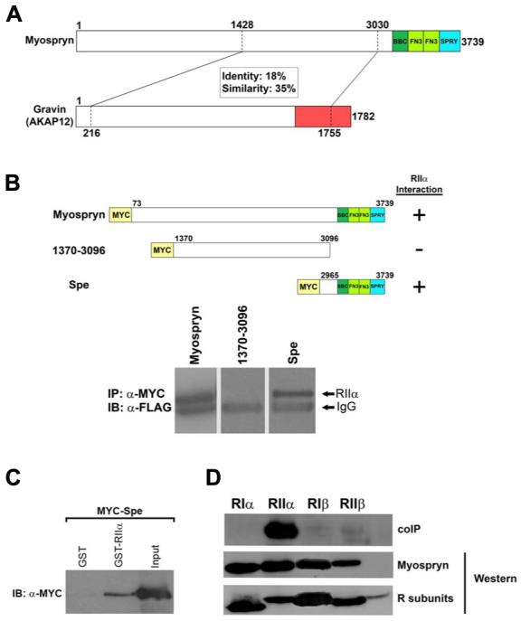
Recently we identified a novel target gene of MEF2A named myospryn that encodes a large, muscle-specific, costamere-restricted alpha-actinin binding protein. Myospryn belongs to the tripartite motif (TRIM) superfamily of proteins and was independently identified as a dysbindin-interacting protein. Dysbindin is associated with alpha-dystrobrevin, a component of the dystrophin-glycoprotein complex (DGC) in muscle. Apart from these initial findings little else is known regarding the potential function of myospryn in striated muscle. Here we reveal that myospryn is an anchoring protein for protein kinase A (PKA) (or AKAP) whose closest homolog is AKAP12, also known as gravin/AKAP250/SSeCKS. We demonstrate that myospryn co-localizes with RII alpha, a type II regulatory subunit of PKA, at the peripheral Z-disc/costameric region in striated muscle. Myospryn interacts with RII alpha and this scaffolding function has been evolutionarily conserved as the zebrafish ortholog also interacts with PKA. Moreover, myospryn serves as a substrate for PKA. These findings point to localized PKA signaling at the muscle costamere.


Figures

Figure 1
Figure 1
Sequence similarity between Myospryn and Gravin. A, Bioinformatics analysis revealed that Myospryn (amino acids 1,428 - 3,030) shares 18% amino acid identity and 35% positive similarity with the PKA anchoring protein Gravin/AKAP12 (amino acids 216 - 1,755). The RIIα̣ interaction domain for Gravin is located at amino acids 1540 through 1553. B, Coimmunoprecipitation of RIIα̣ with the C-terminus of Myospryn. Full length Myospryn is shown to interact with RIIα, while an N-terminal construct containing amino acids 1370 through 3096 is unable to interact. A C-terminal fragment containing amino acids 2965 through 3739 (Spe) is capable of interacting with RIIα. Immunoblot of Myc-immunoprecipitated constructs detecting the presence of FLAG-RIIα. The IgG band is indicated. C, GST pulldown assay. Protein extracts from COS cells transfected with Myc-Spe was added to bacterially expressed GST or GST-RIIα bound to glutathione sepharose beads. Reactions were subjected to SDS-PAGE and immunoblotted using an anti-Myc antibody. D, PKA regulatory subunit specificity for Myospryn. Coimmunoprecipitation of the four regulatory subunits demonstrates that Myospryn is an RIIα-selective AKAP.
Figure 2
Figure 2
Myospryn and RIIα interact in a cellular environment. A, RIIα and NLS-RIIα were transfected into COS cells and immunostained using an anti-RII antibody. RIIα exhibits a cytoplasmic/perinuclear localization whereas NLS-RIIα is shuttled into the nucleus. DAPI staining indicates the location of the nuclei. Arrows demonstrate representative cells. B, COS cells transfected with Myc-Spe alone or cotransfected with NLS-RIIα and Myc-Spe. Myc-Spe alone exhibits a cytoplasmic staining pattern. Cotransfection with NLS-RIIα, Myc-Spe is shuttled into the nucleus demonstrating an in vivo association with this regulatory subunit of PKA. DAPI staining indicates the location of the nuclei. C, Myospryn precipitates PKA from COS cells. COS cells were transfected with pCDNA3-Myc or Myc-Spe. Protein extracts were immunoprecipitated using the anti-MYC antibody and immunoblotted for endogenous PKA using antibodies directed against catalytic subunit (PKA-C, BD Transduction Laboratories). Protein extracts loaded and blotted with the anti-PKA-C antibody detected the presence of similar amounts of PKA-C in both samples.
Figure 3
Figure 3
Co-localization of Myospryn and RIIα in striated muscle. A, Double immunohistochemistry on longitudinal sections from hindlimb muscle. Upper panel, muscle sections using anti-Myospryn antibodies shows a striated pattern of expression (red) as previously demonstrated. Middle panel, anti-RII antibodies reveal a striated pattern of expression (middle) as previously reported. Bottom panel, when the above two images are superimposed a yellow striated signal is readily apparent demonstrating co-localization of Myospryn and RIIα. B, Immunohistochemistry on transverse skeletal muscle. Double immunohistochemistry demonstrates co-localization of myospryn and RII along periphery of skeletal muscle fibers.
Figure 4
Figure 4
Mapping of the RIIα-binding site of Myospryn. A, Coimmunoprecipitation demonstrating the identification of an RIIα interaction domain. Upper panel, Progressive Myc-tagged C-terminal myospryn deletions were cotransfected with FLAG-tagged RIIα, and immunoprecipitated with the Myc antibody and immunoblotted with the FLAG antibody detecting the presence of RIIα. Bottom panel, protein extracts loaded and immunoblotted using the anti-Myc antibody detecting proper expression of the myospryn constructs. Protein extracts loaded and blotted with the anti-FLAG antibody detected similar expression levels of the RIIα construct (not shown). B, Identification of a third amphipathic helix in myospryn. Removal of two amphipathic helices fails to disrupt interaction with RIIα. Additional deletion constructs reveal the presence of a third amphipathic helix. C, Mutational analysis of three amphipathic helices in myospryn. Introduction of point mutations in the proposed amphipathic helices 1 (H1) and H2. Replacement of leucine 3197 with a proline contained within H1 results in the inability of that construct to interact with RIIα, demonstrated by Co-IP. Replacement of leucine 3309 with a proline within H2 results in an inability of that construct to interact with RIIα as well. Replacement of cysteine with a proline in H3 severely diminishes interaction with RIIα. D, Helical wheel representation of the three amphipathic helices. Shown is a clustering of hydrophobic residues (shaded) on one face of the helix and hydrophilic residues (unshaded) on the other face, a feature characteristic of an amphipathic helix which confers the ability of an AKAP to interact with PKA.
Figure 5
Figure 5
A, Schematic diagram of select Myc-tagged Myospryn constructs expressed in COS cells to map the RIIα-binding domain. Interactions results are shown at right (+, positive interaction; -, negative interaction). B, Schematic depiction of the three PKA anchoring motifs in relation to the TRIM region of Myospryn. The H1 helix is localized immediately upstream of the TRIM region; H2 is found within the B-box coiled coil (BBC) domain; and H3 is situated within the first fibronectin 3 repeat (FN3).
Figure 6
Figure 6
A, Sequence alignment of the three PKA anchoring motifs. The C-terminal myospryn fragment Spe possesses significant sequence homology to the Zebrafish (Danio rerio) myospryn ortholog (amino acids 894 and 1161). This region in zebrafish myospryn harbors three proposed RIIα interacting domains. B, Co-immunoprecipitation of zebrafish myospryn and RIIα. Like mouse myospryn, zebrafish myospryn is capable of immunoprecipitating RIIα, demonstrating an evolutionary conservation AKAP function for myospryn.
Figure 7
Figure 7
Myospryn is a substrate for PKA. A, Schematic of the N-terminal fragment of myospryn (amino acids 73-743) shows three consensus PKA phosphorylation sites between amino acids 138-158. B, The putative PKA phosphorylation sites are phosphorylated by PKA in vitro. The N-terminal myospryn fragment was subjected to an in vitro kinase assay using recombinant catalytic subunit of PKA. Deletion of all three sites from this myospryn fragment is unable to be phosphorylated by PKA in vitro. C, The N-terminal fragment of myospryn containing the PKA consensus sites was incubated in the kinase assay buffer without PKA enzyme demonstrating that other endogenous kinases were not responsible for the phosphorylation. Incubation with recombinant PKA and the PKA-specific peptide inhibitor, PKI, resulted in a loss of phosphorylation of the N-terminal fragment demonstrating specificity for PKA. For all of the above experiments protein expression of the wildtype and mutant proteins was confirmed by blotting protein extracts with the anti-FLAG antibody.

Similar articles

Cited by

References

    1. Black BL, Olson EN. Transcriptional control of muscle development by myocyte enhancer factor-2 (MEF2) proteins. Annu Rev Cell Dev Biol. 1998;14:167–96. - PubMed
    1. McKinsey TA, Zhang CL, Olson EN. MEF2: a calcium-dependent regulator of cell division, differentiation and death. Trends Biochem Sci. 2002;27(1):40–7. - PubMed
    1. Blais A, Tsikitis M, Acosta-Alvear D, Sharan R, Kluger Y, Dynlacht BD. An initial blueprint for myogenic differentiation. Genes Dev. 2005;19(5):553–69. - PMC - PubMed
    1. Paris J, Virtanen C, Lu Z, Takahashi M. Identification of MEF2-regulated genes during muscle differentiation. Physiol Genomics. 2004;20(1):143–51. - PubMed
    1. Junion G, Jagla T, Duplant S, Tapin R, Da Ponte JP, Jagla K. Mapping Dmef2-binding regulatory modules by using a ChIP-enriched in silico targets approach. Proc Natl Acad Sci U S A. 2005;102(51):18479–84. - PMC - PubMed

Publication types

MeSH terms

Substances