. 2007 Jun;7(6):1757-63.
doi: 10.1021/nl070828k. Epub 2007 May 15.

Six-helix and eight-helix DNA nanotubes assembled from half-tubes

Affiliations

Six-helix and eight-helix DNA nanotubes assembled from half-tubes

Akinori Kuzuya et al. Nano Lett. 2007 Jun.

Abstract

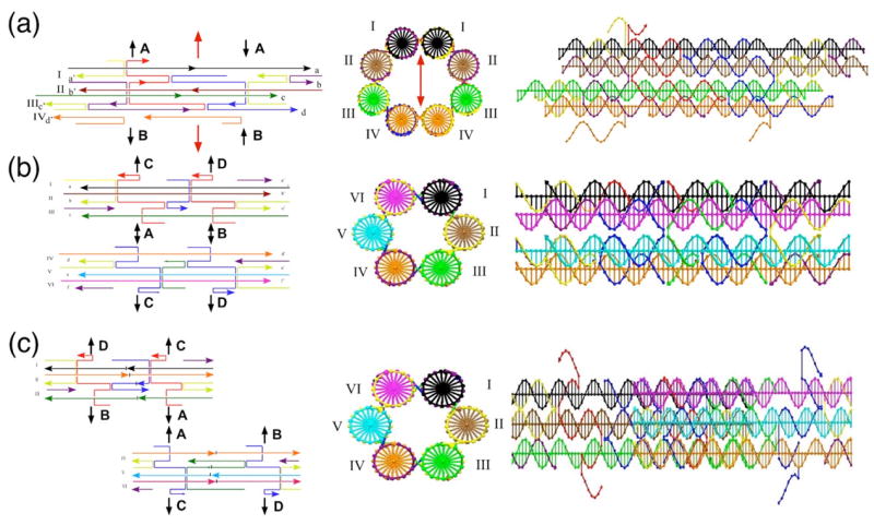
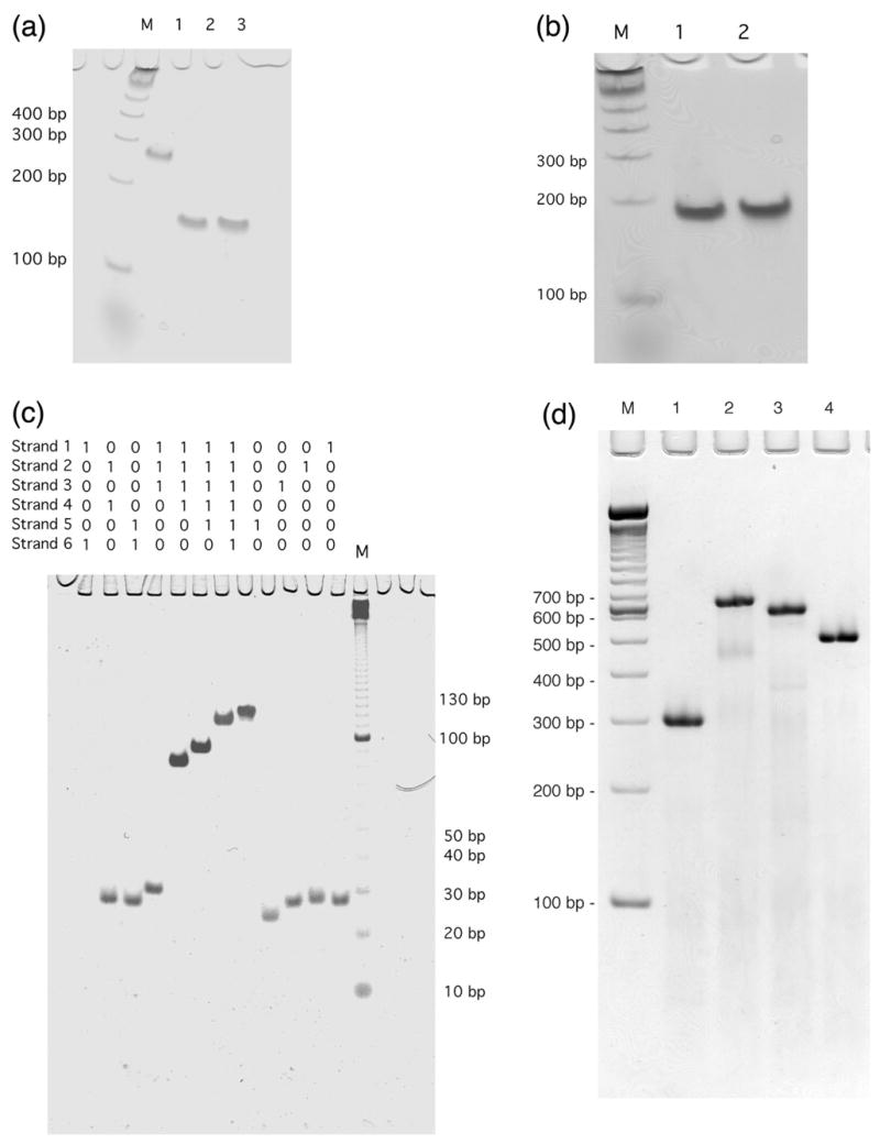
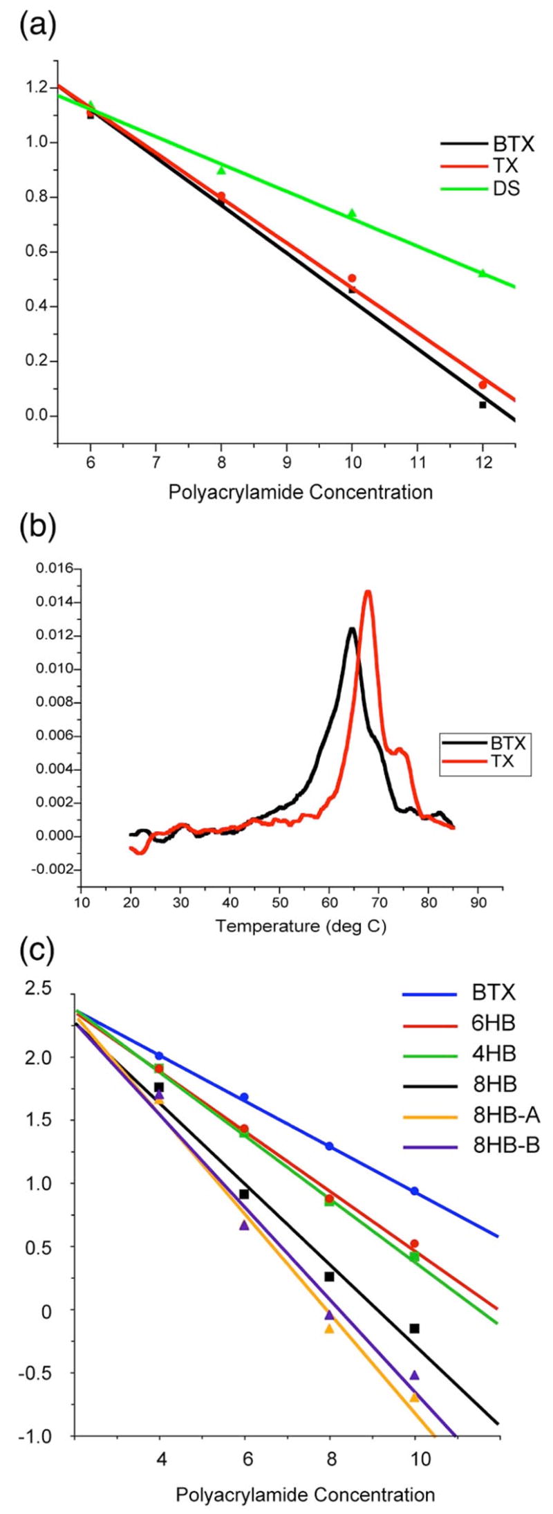
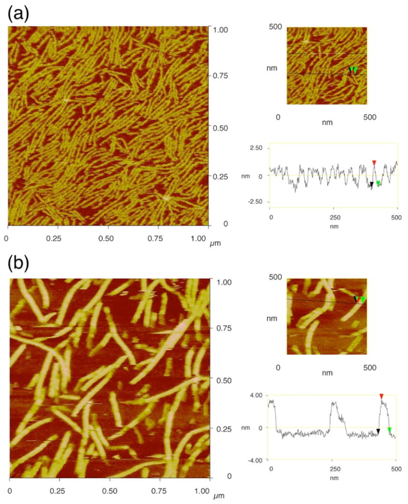
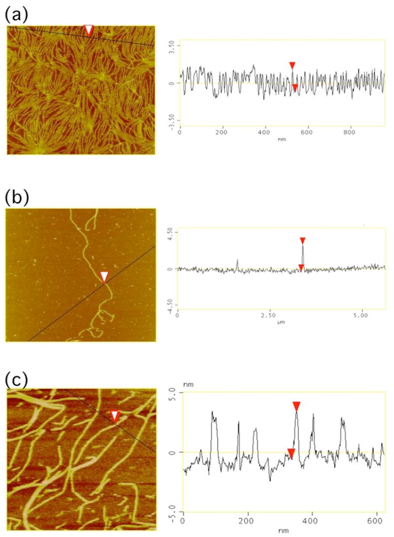
DNA nanotubes are cylinder-like structures formed from DNA double-helical molecules whose helix axes are fused at least twice by crossovers. It is potentially useful to use such tubes as sheaths around rodlike species that arise in biological systems and in nanotechnology. It seems easiest to obtain such sheathing by joining two or more components around an object rather than attempting to thread the object through a cavity in the tube. We report two examples of tubes containing a specific number of helices that are assembled from half-tube components. These tubes are a six-helix bundle and an eight-helix bundle, constructed respectively from two bent triple-crossover (BTX) molecules and from two four-helix arched motifs. Both species contain single strands in one molecule that are missing in its mate. The six-helix bundle is formed from two different BTX molecules, whereas the eight-helix species is a closed cyclic dimer of the same molecule. We demonstrate the formation of these species by gel electrophoresis, and we examine their arrangement into long one-dimensional arrays by means of atomic force microscopy.


Figures

Figure 1
Figure 1
Schematic Drawings of DNA Half-Tube Molecules. At the left of each panel is a line drawing indicating the structure of the half-tube molecules and the ways in which the molecules connect to each other. These are indicated by letters and arrows that connect to the same letters and arrows. Lateral connections are shown as gaps and unpaired strands. In the center of the panels the cross-sections of the complete tubes are shown schematically, and the helices are labeled with Roman numerals. To the right is a view with the helix and tube axes horizontal. (a) The 4HB half-tube molecule. The central portion shows an elliptical cross-section for the 8HB molecule. The dyad axis relating it to itself is drawn vertically as a red double-headed arrow. Only the 4HB molecule is shown at right. (b) The BTX molecules with similar phasing. (c) The BTX molecules phased half a length apart.
Figure 2
Figure 2
Non-Denaturing Gels Demonstrating Formation of Half-Tube Components. (a) Blunt-ended versions of the BTX molecules phased opposite each other. A marker lane containing 100 nt fragments is labeled M. Lanes 2 and 3 contain the two components and lane 1 shows their cohesion as a clean band. (b) Blunt-ended versions of the molecules that are offset by a half length. Both lanes contain a well-formed single band. (c) The subcomponents of a blunt-ended BTX molecule. The contents of individual lanes are indicated above them, with a ‘1’ indicating the presence of the component. A 10 nt fragment marker lane is at right. (d) Formation of the 8HB from the 4HB molecule. A 100 nt marker lane is labeled ‘M’. Lane 1 contains the individual 4HB molecule without lateral or terminal single strands. Lane 2 contains two molecules joined only at point A (8HB-A), and lane 3 contains two molecules joined only at point B (8HB-B). Both lanes contain various breakdown products. Lane 4 contains the complete 8HB without sticky ends. It is cleanly formed.
Figure 3
Figure 3
Characterization of BTX and 4HB molecules. (a) Ferguson plots comparing BTX to a conventional planar TX molecule of the same length. A linear duplex molecule (slope = 0.100) is shown for comparison. The BTX molecule is similar, but its slope (0.175) is slightly higher than the planar molecule (0.165). (b) Differential melting plots of the BTX and TX molecules. Note that BTX melts at slightly lower temperatures. (c) Ferguson plots of the species used here. Slopes are BTX (0.180), 6HB (0.235), 4HB (0.251), 8HB (0.319), 8HB-A (0.396), 8HB-B (0.369).
Figure 4
Figure 4
Atomic Force Micrographs of 4HB Half-Tube arrays and 8HB Nanotubes. Both panels contain an image of 1D arrays on the left and a height-analysis on the right. (a) The 4HB half-tube array. The colored arrows on the line indicate the position of the height analysis. (b) The 8HB nanotubes. Note that the tubes are much higher for the 8HB species.
Figure 5
Figure 5
BTX arrays and 6HB Nanotubes. The same conventions apply as in Figure 4, except that the scan line for height analysis is part of the image on the left side of the panel. (a) Long BTX arrays are shown; the image is 950 nanometers square. (b) A 6HB nanotube formed from a pair of BTX molecules phased opposite each other; the image is 5 microns square. (c) 6HB nanotubes formed from the overlapping motif, with BTX molecules phased half a length apart; the image is 820 nanometers square.

References

    1. Holliday R. Genet Res. 1964;5:282–304.
    1. Fu TJ, Seeman NC. Biochem. 1993;32:3211–3220. - PubMed
    1. Sa-Ardyen P, Vologodskii AV, Seeman NC. Biophys J. 2003;84:3829–3837. - PMC - PubMed
    1. Winfree E, Liu F, Wenzler LA, Seeman NC. Nature. 1998;394:539–544. - PubMed
    1. Rothemund PWK, Papadakis N, Winfree E. PLOS Biology. 2004;2:2041–2053. - PMC - PubMed

Publication types