. 2007 May;64(4):923-37.
doi: 10.1111/j.1365-2958.2007.05694.x.

The novel ER membrane protein PRO41 is essential for sexual development in the filamentous fungus Sordaria macrospora

Affiliations

The novel ER membrane protein PRO41 is essential for sexual development in the filamentous fungus Sordaria macrospora

Minou Nowrousian et al. Mol Microbiol. 2007 May.

Abstract

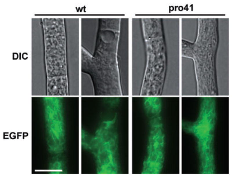
The filamentous fungus Sordaria macrospora develops complex fruiting bodies (perithecia) to propagate its sexual spores. Here, we present an analysis of the sterile mutant pro41 that is unable to produce mature fruiting bodies. The mutant carries a deletion of 4 kb and is complemented by the pro41 open reading frame that is contained within the region deleted in the mutant. In silico analyses predict PRO41 to be an endoplasmic reticulum (ER) membrane protein, and a PRO41-EGFP fusion protein colocalizes with ER-targeted DsRED. Furthermore, Western blot analysis shows that the PRO41-EGFP fusion protein is present in the membrane fraction. A fusion of the predicted N-terminal signal sequence of PRO41 with EGFP is secreted out of the cell, indicating that the signal sequence is functional. pro41 transcript levels are upregulated during sexual development. This increase in transcript levels was not observed in the sterile mutant pro1 that lacks a transcription factor gene. Moreover, microarray analysis of gene expression in the mutants pro1, pro41 and the pro1/41 double mutant showed that pro41 is partly epistatic to pro1. Taken together, these data show that PRO41 is a novel ER membrane protein essential for fruiting body formation in filamentous fungi.


Figures

Fig. 1
Fig. 1. Complementation analysis of the pro41 mutant
A. The S. macrospora cosmids C6 and G2 as well as the N. crassa cosmid pMOcosXG20A10 complement mutant pro41. White arrows, predicted N. crassa open reading frame; black regions, elements that were verified to be present on the S. macrospora cosmids; light grey, smallest complementing region deduced from complementation analysis with cosmids. B. The pro41 open reading frame complements the developmental defects of mutant pro41. White arrows, predicted S. macrospora open reading frames; dark grey bar, region that is deleted in mutant pro41; white bars, complementing clone/fragment; light grey, smallest complementing region; black bars, non-complementing clones; hatched bars, regulatory regions from A. nidulans (gpd promoter, trpC terminator).
Fig. 2
Fig. 2
Multiple alignment of PRO41 orthologues from filamentous fungi. The alignment was created using CLUSTALX (Thompson et al., 1997) with the S. macrospora PRO41 (S. m., emb|AM410183), and sequences from fully sequenced genomes of the following fungi: Nc, Neurospora crassa NCU02767.2; Cg, Chaetomium globosum CHG01547.1; Fg, Fusarium graminearum FG01268.1; Mg, Magnaporthe grisea MGG_09956.5; Ss, Sclerotinia sclerotiorum SS1G_04691.1; An, Aspergillus nidulans AN5458.3; Af, Aspergillus fumigatus Afu6g13340; Ci, Coccidioides immitis CIMG_10237.2; Hc, Histoplasma capsulatum HCAG_05179.1; Sn, Stagonospora nodorum SNOG_11389.1. The A. nidulans, A. fumigatus, H. capsulatum and S. nodorum PRO41 orthologues were manually re-annotated as the predicted proteins are shorter than all other PRO41 orthologues probably due to annotation errors. Sequences were obtained from the Aspergillus fumigatus sequence project (http://www.tigr.org/tdb/e2k1/afu1/) for A. fumigatus and from the genome databases of the Fungal Genome Initiative (http://www.broad.mit.edu/annotation/fungi/fgi/index.html) for all other sequences. Jalview was used to visualize the alignment (Clamp et al., 2004). Amino acid residues conserved in at least nine sequences are given in dark grey, residues conserved in at least seven sequences in medium grey, and in at least five sequences in light grey. The N-terminal signal sequence as predicted by PSORT and SIGNALP (Nakai and Horton, 1999; Bendtsen et al., 2004) is shown by a dark grey bar above the sequence, putative cleavage sites are indicated by black triangles. Transmembrane domains that were predicted by TMPRED (Hofmann and Stoffel, 1993) are shown by light grey bars below the sequences. A putative ER retention signal is indicated by an open box above the sequence. Amino acid identities in percentage of the PRO41 orthologues with each other are given at the end of the alignment.
Fig. 3
Fig. 3. Analysis of the PRO41 protein
A. The predicted PRO41 signal sequence for co-translational insertion into the ER is functional. Constructs for expression of egfp (pEH3), and egfp fused with the signal sequences from ppg1 or pro41 (pSppg1-1 and pSpro41 respectively) were transformed into the wild type. Protein extracts from the culture medium of transformants were separated by SDS-PAGE and analysed for secreted EGFP by immunodetection with an anti-EGFP antibody. B. A PRO41–EGFP fusion protein localizes to the ER in a signal sequence-dependent manner. Mutant pro41 was co-transformed with pE3-5Mr and pDsREDKDEL (left) or with pE3-7Mr and pDsREDKDEL (right). pE3-5Mr expresses full-length pro41 fused to egfp, whereas in pE3-7Mr, the N-terminal signal sequence of pro41 is missing (pro41ΔS). pDsREDKDEL expresses ER-localized dsred. Transformants were analysed by fluorescence microscopy. Scale bar = 10 μm. C and D. A PRO41–EGFP fusion protein localizes to membranes. Mutant pro41 was transformed with pEH3 or pE3-5Mr (plasmids as described in A and B). C. Total protein extract (T) from the mycelia of transformants was separated into a pellet (P) containing the membrane fraction and a supernatant (S) containing the soluble fraction after centrifugation at 100 000 g. Western blot analysis was performed with an anti-GFP antibody. Analyses were performed with three independent transformants each. Results for only one representative transformant are shown. D. Total protein extract from a pro41–egfp-expressing transformant as described in C was subjected to differential centrifugation. Supernatants (S) and pellets (P) of the consecutive centrifugation steps (9k, 9000 g; 20k, 20 000 g; 40k, 40 000 g; 100k, 100 000 g) were separated by SDS-PAGE, and Western blot analysis was performed with an anti-GFP antibody and or anti-TIM23 antibody respectively.
Fig. 4
Fig. 4
ER morphology is normal in mutant pro41. Plasmid pEGFPKDEL expressing ER-localized EGFP was transformed into the S. macrospora wild type and mutant pro41. Vegetative hyphae of transformants were analysed by fluorescence microscopy. The net-like structures representing the ER are similar in mutant pro41 and the wild type. Scale bar = 10 μm.
Fig. 5
Fig. 5
Expression analysis of the pro41 gene. Quantitative real-time PCR analysis of pro41 expression in the developmental mutants pro1, pro11 and pro22, as well as in the wild type growing vegetatively. Comparisons were against the wild type undergoing sexual development. Values given are mean expression ratios and standard deviations from at least two biological replicates (n = 2–6). Black bars indicate that pro41 is significantly downregulated under this condition (P < 0.05, calculated with REST) (Pfaffl et al., 2002).
Fig. 6
Fig. 6. Molecular epistasis analysis of pro1 and pro41 by microarray hybridizations. Gene expression in the wild type growing vegetatively as well as mutants pro1, pro41 and pro1/41 was compared with the wild type undergoing sexual development. Microarray data were analysed using the Bioconductor package ‘limma’ and correspondence analysis (COA) was performed with the package ‘made4’ (Fellenberg et al., 2001; Smyth, 2004; Culhane et al., 2005)
A. Hierarchical clustering of targets (in this case the different strains pro1, pro41, pro1/41, and wt vegetatively) according to the similarity of gene expression patterns. B. COA of microarray data from the wild type and developmental mutants. Gene expression in the wild type growing vegetatively as well as mutants pro1, pro41 and pro1/41 were compared with the wild type undergoing sexual development. The 500 most differentially expressed genes in any of these comparisons were used for COA. A list of the genes used in this analysis is given in Fig. S3. The biplot shows genes as black dots, and the five most differentially expressed genes at both ends of each axis are labelled in blue, red, green and orange with the number of the corresponding N. crassa open reading frame indicated. Targets are given as grey ovals. Genes that show strong expression in a certain strain are located in the direction determined by the target of this strain. The farther away from the centre, the more pronounced is the association of the genes with that strain. Genes that are downregulated in this phase appear on the opposite site of the centroid. C. Model for the genetic interaction between pro1 and pro41 in fruiting body development.

Comment in

Similar articles

Cited by

References

    1. Altschul SF, Madden TL, Schaffer AA, Zhang J, Zhang Z, Miller W, Lipman DJ. Gapped BLAST and PSI-BLAST: a new generation of protein database search programs. Nucleic Acids Res. 1997;25:3389–3402. - PMC - PubMed
    1. Beers J, Glerum DM, Tzagoloff A. Purification, characterization, and localization of yeast Cox17p, a mitochondrial copper shuttle. J Biol Chem. 1997;272:33191–33196. - PubMed
    1. Bendtsen JD, Nielsen H, von Heijne G, Brunak S. Improved prediction of signal peptides: SignalP 3.0. J Mol Biol. 2004;340:783–795. - PubMed
    1. Bergeron JJ, Rachubinski RA, Sikstrom RA, Posner BI, Paiement J. Galactose transfer to endogenous acceptors within Golgi fractions of rat liver. J Cell Biol. 1982;92:139–146. - PMC - PubMed
    1. Berteaux-Lecellier V, Picard M, Thompson-Coffe C, Zickler D, Panvier-Adoutte A, Simonet JM. A nonmammalian homolog of the PAF1 gene (Zellweger syndrome) discovered as a gene involved in caryogamy in the fungus Podospora anserina. Cell. 1995;81:1043–1051. - PubMed

Publication types

MeSH terms