. 2007 Jul;27(14):5201-13.
doi: 10.1128/MCB.02237-06. Epub 2007 May 14.

Transcriptional repressor erf determines extraembryonic ectoderm differentiation

Affiliations

Transcriptional repressor erf determines extraembryonic ectoderm differentiation

Chara Papadaki et al. Mol Cell Biol. 2007 Jul.

Abstract

Extraembryonic ectoderm differentiation and chorioallantoic attachment are fibroblast growth factor (FGF)- and transforming growth factor beta-regulated processes that are the first steps in the development of the placenta labyrinth and the establishment of the fetal-maternal circulation in the developing embryo. Only a small number of genes have been demonstrated to be important in trophoblast stem cell differentiation. Erf is a ubiquitously expressed Erk-regulated, ets domain transcriptional repressor expressed throughout embryonic development and adulthood. However, in the developing placenta, after 7.5 days postcoitum (dpc) its expression is restricted to the extraembryonic ectoderm, and its expression is restricted after 9.5 dpc in a subpopulation of labyrinth cells. Homozygous deletion of Erf in mice leads to a block of chorionic cell differentiation before chorioallantoic attachment, resulting in a persisting chorion layer, a persisting ectoplacental cone cavity, failure of chorioallantoic attachment, and absence of labyrinth. These defects result in embryo death by 10.5 dpc. Trophoblast stem cell lines derived from Erf(dl1/dl1) knockout blastocysts exhibit delayed differentiation and decreased expression of spongiotrophoblast markers, consistent with the persisting chorion layer, the expanded giant cell layer, and the diminished spongiotrophoblast layer observed in vivo. Our data suggest that attenuation of FGF/Erk signaling and consecutive Erf nuclear localization and function is required for extraembryonic ectoderm differentiation, ectoplacental cone cavity closure, and chorioallantoic attachment.


Figures

FIG. 1.
FIG. 1.
Targeted disruption of the Erf gene. (A) Schematic representation of the Erf targeting strategy. (1) Genomic organization of the murine Erf gene. Exons are indicated by black boxes and are numbered 1 to 4. (2) The structure of the targeting vector. The AvrII-BamHI fragment was replaced by the pMC1neo cassette (gray box) flanked by loxP sites (black triangles). The arrow above indicates the orientation of the neo gene transcription. Dashed lines outline the Erf genomic sequences incorporated into the targeting construct. The herpes simplex virus thymidine kinase cassette (white box) was placed at both ends of the targeting vector for negative selection. (3) The predicted structure of the targeted Erf locus (Erfdl1-neo) following homologous recombination of the targeting vector in ES cells. (4) The predicted structure of the targeted Erf locus (Erfdl1) following Cre-mediated excision. Small arrows indicate the primers (p1, p2, and p3) used for genotyping by PCR. Boldface lines indicate the position of probes used for the Southern analysis. The sizes of the expected restriction fragments are also indicated. Restriction enzymes are the following: A, AvrII; B, BamHI; E, EcoRI; N, NotI; P, PvuII; S, SacI; X, XbaI. (B) Homologous recombination was verified by Southern analysis. Genomic DNA from control ES cells and two independently targeted ES clones, no. 130 and no. 169, was digested with EcoRI and hybridized to the radiolabeled 3′ external (3′ext) probe located outside of the targeting vector. The 14-kb fragment corresponding to the wild-type (wt) allele and the 5-kb fragment corresponding to the Erfdl1-neo allele are indicated. (C) Hybridization of genomic EcoRI fragments with the 5′ext probe. The 14-kb wild-type fragment and the 9-kb Erfdl1-neo fragment are indicated. (D) Southern blot analysis of genomic DNA from F1 mice. DNA was digested with BamHI and hybridized to the radiolabeled internal (int) probe. Cre-recombined heterozygous mutant mice (dl1) were identified by the smaller 7-kb fragment, compared to the 8.3-kb mutant fragment of the nonrecombined allele (dl1-neo). The 3-kb band corresponds to the wild-type allele. (E) Southern blot analysis of genomic DNA derived from 9.5-dpc embryos of Erfdl1/+ heterozygous mice intercross. The 3-kb band corresponds to the wild-type allele, and the 7-kb band corresponds to the Erfdl1 allele. (F) PCR analysis of genomic DNA derived from visceral yolk sacs of 9.5-dpc embryos from Erfdl1/+ intercrosses using p1 and p3 primers. The wild-type allele is identified by the 1-kb band, and the Erfdl1 allele is identified by the 300-bp band. (G) Northern analysis of total RNA extracted from wild-type, heterozygous, and homozygous 9.5-dpc embryos, free of extraembryonic tissues, hybridized with the radiolabeled int probe (upper panel). Loading and integrity of RNA was assessed by ethidium bromide staining of the 28S and 18S rRNA in the gel (lower panel).
FIG. 2.
FIG. 2.
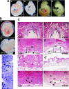
Phenotype of the Erf-deficient mouse embryos. (A) Gross morphology of 9.5-dpc wild-type (a) and Erfdl1/dl1 (b) embryos and 10.5-dpc wild-type (c) and Erfdl1/dl1 (d) embryos. (B) Yolk sac morphology of a wild-type (a) and an Erfdl1/dl1 (b) embryo at 9.5 dpc. A highly organized vascular network is formed in wild-type yolk sacs, whereas in Erfdl1/dl1 embryos the yolk sacs are pale and exhibit only the primary capillary plexus without further reorganization. Whole-mount staining of wild-type (c) and Erfdl1/dl1 (d) yolk sacs with hematoxylin are also shown. Large collecting vessels are formed in both wild-type and Erfdl1/dl1 yolk sacs (arrowheads), but in Erfdl1/dl1 they are enlarged and do not form secondary branches (arrows) as in the wild-type littermates. (C) Hematoxylin- and eosin-stained paraffin sections from wild-type (a, c, c′, and e) and Erfdl1/dl1 (b, d, d′, and f) littermates. At 8.5 dpc the allantois is attached throughout the chorion layer in the wild-type placentas (panel a, arrowheads) but not in Erfdl1/dl1 placentas, where the ectoplacemmtal cone cavity is still open (panel b, asterisk). At 9.5 dpc allantoic blood vessels invade the chorion layer in wild-type placentas (arrowheads in panels c, c′), but in the Erfdl1/dl1 placentas the chorion retains a compact structure, the labyrinth is totally absent (d′), and the ectoplacental cone cavity is still present (panel d, asterisk). At 10.5 dpc in wild-type placentas, maternal and embryonic blood vessels (panel e, arrow and arrowhead, respectively) intermingle and come to close apposition. In contrast, Erfdl1/dl1 placentas appear degenerative, with extensive hemorrhagic sites (panel f, arrows). al, allantois; ch, chorion; dec, decidua; gc, giant cells; lb, labyrinth; sp, spongiotrophoblast; uc, umbilical cord. Yellow dotted lines indicate the boundaries of the placenta.
FIG. 3.
FIG. 3.
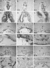
Expression of Erf in the developing placenta. In situ hybridization of Erf mRNA in sagittal sections of wild-type placentas from 6.5 to 15.5 dpc using a 1-kb riboprobe derived from Erf exon 4. (A to C) Through early placenta development (6.5 to 8.0 dpc) Erf mRNA is detected in the ExE. (D) At 8.5 dpc Erf mRNA is detected throughout the chorionic ectoderm (ChE). (E) By 9.5 dpc Erf mRNA persists in the chorion (ch). It is also detected in some trophoblast cells in the developing labyrinth. (F and G) At 10.5 to 12.5 dpc Erf transcripts are present in the chorion and in the labyrinthine trophoblast cells. (H and I) At 13.5 dpc Erf mRNA expression in the chorion and in labyrinthine trophoblasts is decreased and disappears after 15.5 dpc. (a to i) Magnification of the indicated areas (boxes) of panels A through I. al, allantois; e, embryo; epc, ectoplacental cone; lb, labyrinth; sp, spongiotrophoblast; uc, umbilical cord. Dotted lines indicate the boundaries of the placenta.
FIG. 4.
FIG. 4.
Failure of chorioallantoic attachment in Erfdl1/dl1 embryos. (A) Confocal microscopy images from sagittal sections of 8.5-dpc (a and b) and 9.5-dpc (c and d) placentas. Freshly frozen placentas were stained with antibodies against the allantoic protein VCAM-1 (red) and the chorionic protein integrin α4 (green). Nuclei were stained with TOPRO-3 (blue). (a) In 8.5-dpc wild-type (WT) placentas, attachment of allantoic mesothelium with chorionic mesoderm throughout the chorion layer is evidenced by the yellow color (arrowheads). (b) Both VCAM1 and integrin α4 are expressed in Erfdl1/dl1 placentas. The mesothelial surface of allantois approaches the chorionic mesoderm (arrows) but fails to fuse and spread throughout the chorionic layer. (c) In 9.5-dpc wild-type placentas, chorioallantoic fusing surfaces break down, enabling the juxtaposition of allantoic vasculature and chorion layer, resulting in the penetration of allantoic endothelium to the chorion, indicated by the VCAM1 staining at both sides of the chorion layer (arrowheads). (d) In Erfdl1/dl1 placentas, the proximity of allantois can be seen throughout the chorionic mesoderm, but there is no penetration of allantoic mesothelium or even contact of the two layers, as indicated by the lack of yellow color. Yellow dotted lines indicate the boundaries of the placenta. (B) PECAM staining (red) in wild-type and Erfdl1/dl1 placentas from 9.5 dpc. Nuclei were stained blue with TOPRO-3. The yellow lines in the right panel indicate the chorion layer in Erfdl1/dl1 placentas. al, alantois; lb, labyrinth; ch, chorion; sp, spongoid layer; gc, giant cells; fbv, fetal blood vessels.
FIG. 5.
FIG. 5.
Expression of cell type-specific markers in the developing placenta. (A) In situ hybridization was performed on frozen sections from 8.5-dpc wild-type (WT) (a to d) and Erfdl1/dl1 (e to h) placentas with antisense RNA probes for Pl1, Tpbpa, Gcm1, and Errβ. Expression of Pl1 is unaffected in Erfdl1/dl1 placentas (a and e). Expression of Tpbpa is reduced in Erfdl1/dl1 placentas (b and f). Gcm1, which is expressed in a subset of chorionic trophoblast cells in wild-type placentas, is absent from Erfdl1/dl1 placentas (c and g). Errβ is expressed (arrowheads) at the margins of the chorion in wild-type placentas, while in Erfdl1/dl1 placentas (h) it is detected throughout the chorion (d and h). (B) In situ hybridization on sections from 9.5-dpc wild-type (a to d) and Erfdl1/dl1 (e to h) placentas with probes for Pl1, Tpbpa, Gcm1, and Tef5. Expression of Pl1 in Erfdl1/dl1 placentas reveals an expansion of trophoblast giant cell layer (e and e′). Expression of Tpbpa shows a reduced spongiotrophoblast cell layer in Erfdl1/dl1 placentas (f and f′). Gcm1 is expressed in syncytiotrophoblasts of the labyrinthine layer and in chorionic trophoblast cells in wild-type placentas (c and c′) but is not detectable in Erfdl1/dl1 placentas (g and g′). Tef5 is expressed in syncytiotrophoblasts and in labyrinthine trophoblasts in wild-type placentas (d and d′) but is not present in Erfdl1/dl1 placentas (h and h′). (a′ to h′) Magnification of the indicated regions (white rectangles) from panels a through h, respectively. (C) In situ hybridization on sections from 10.5-dpc placentas as described for panel B. The giant cell layer remains expanded (a and e), the trophoblast cell layer is disorganized (b and f), and the sycytiotrophoblast cells (c, g, d, and h) are missing in Erfdl1/dl1 animals. dec, decidual; al, allantois; lb, labyrinth; sp, spongiotrophoblasts; gc, giant cells. Dotted lines indicate the boundaries of the placenta.
FIG. 6.
FIG. 6.
Gcm1 expression in the developing placenta at 8.0 dpc. In situ hybridization was performed on frozen sections from 8.0-dpc wild-type (upper panel) and Erfdl1/dl1 (lower panel) placentas with Gcm1 (left panels) and Errβ (right panels) antisense RNA probes. The arrows indicate points of Gcm1 expression. dec, decidual; ch, chorion; WT, wild type. Dotted lines indicate the boundaries of the placenta.
FIG. 7.
FIG. 7.
Gene expression in differentiating TSCs. (A) RNA from wild-type (WT) TSC under proliferation conditions at the indicated cell densities (day 0) and the indicated times (differentiation [Diff.] day) in differentiation media was analyzed by Northern blotting for the expression of Erf, the TSC marker Eomes, the giant cell marker Pl1, and GAPDH as a loading control. (B) The level of Erf mRNA during TSC differentiation was determined by QPCR and compared to the Cph mRNA levels in the same sample. The average of two independent wild-type cell lines is shown. (C) Protein extracts from differentiating wild-type TSCs were analyzed by immunoblotting to determine the levels of total (upper panel) and activated (lower panel) Erks. (D) The mRNA levels of Eomes, Cdx2, and Errβ from wild-type (filled symbols) and Erfdl1/dl1 (open symbols) TSCs were analyzed by QPCR and are presented as fractions of the Cph mRNA value in each sample. The graph presents the average of four Erfdl1/dl1 cell lines and two wild-type cell lines. (E) The values of the Erfdl1/dl1 samples from panel D were divided by the corresponding values of the wild-type samples to determine differences in TSC marker expression. (F) As described for panel E, the average values of the Tpbpa levels from the four Erfdl1/dl1 and the two wild-type TSC cell lines were divided to determine the differential expression of Tpbpa during TSC differentiation. Stars in panels E and F indicate statistically significant differences, with P values between 0.01 and 0.05.
FIG. 8.
FIG. 8.
Model for the role of Erf in TSC differentiation. Erf may affect the proper initial commitment of TSCs. At a later stage elevated levels of Erf in the absence of Erk activity are required for the differentiation of chorion diploid cells to terminally differentiated syncytiotrophoblasts (Syn.Tr.), ectoplacental cone cavity closure, and chorioallantic attachment. Ch-TSC, chorion TSC.

Similar articles

Cited by

References

    1. Anson-Cartwright, L., K. Dawson, D. Holmyard, S. J. Fisher, R. A. Lazzarini, and J. C. Cross. 2000. The glial cells missing-1 protein is essential for branching morphogenesis in the chorioallantoic placenta. Nat. Genet. 25:311-314. - PubMed
    1. Arman, E., R. Haffner-Krausz, Y. Chen, J. K. Heath, and P. Lonai. 1998. Targeted disruption of fibroblast growth factor (FGF) receptor 2 suggests a role for FGF signaling in pregastrulation mammalian development. Proc. Natl. Acad. Sci. USA 95:5082-5087. - PMC - PubMed
    1. Bartel, F. O., T. Higuchi, and D. D. Spyropoulos. 2000. Mouse models in the study of the Ets family of transcription factors. Oncogene 19:6443-6454. - PubMed
    1. Basyuk, E., J. C. Cross, J. Corbin, H. Nakayama, P. Hunter, B. Nait-Oumesmar, and R. A. Lazzarini. 1999. Murine Gcm1 gene is expressed in a subset of placental trophoblast cells. Dev. Dyn. 214:303-311. - PubMed
    1. Chen, C., W. Ouyang, V. Grigura, Q. Zhou, K. Carnes, H. Lim, G. Q. Zhao, S. Arber, N. Kurpios, T. L. Murphy, A. M. Cheng, J. A. Hassell, V. Chandrashekar, M. C. Hofmann, R. A. Hess, and K. M. Murphy. 2005. ERM is required for transcriptional control of the spermatogonial stem cell niche. Nature 436:1030-1034. - PMC - PubMed

Publication types

MeSH terms