. 2007 Jul;27(14):5235-45.
doi: 10.1128/MCB.01645-06. Epub 2007 May 14.

Phosphorylation of Bcl10 negatively regulates T-cell receptor-mediated NF-kappaB activation

Affiliations

Phosphorylation of Bcl10 negatively regulates T-cell receptor-mediated NF-kappaB activation

Hu Zeng et al. Mol Cell Biol. 2007 Jul.

Abstract

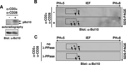
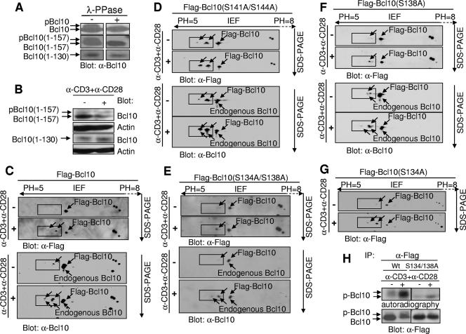
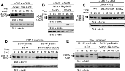
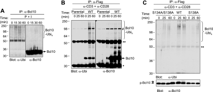
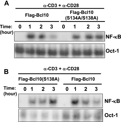
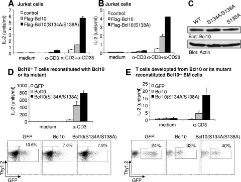
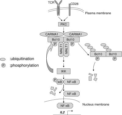
Bcl10 (B-cell lymphoma 10) is an adaptor protein comprised of an N-terminal caspase recruitment domain and a C-terminal serine/threonine-rich domain. Bcl10 plays a critical role in antigen receptor-mediated NF-kappaB activation and lymphocyte development and functions. Our current study has discovered that T-cell activation induced monophosphorylation and biphosphorylation of Bcl10 and has identified S138 within Bcl10 as one of the T-cell receptor-induced phosphorylation sites. Alteration of S138 to an alanine residue impaired T-cell activation-induced ubiquitination and subsequent degradation of Bcl10, ultimately resulting in prolongation of TCR-mediated NF-kappaB activation and enhancement of interleukin-2 production. Taken together, our findings demonstrate that phosphorylation of Bcl10 at S138 down-regulates Bcl10 protein levels and thus negatively regulates T-cell receptor-mediated NF-kappaB activation.


Figures

FIG. 1.
FIG. 1.
Bcl10 is monophosphorylated and biphosphorylated following TCR stimulation. (A) Bcl10 is phosphorylated following TCR engagement in Jurkat cells. Jurkat cells were labeled with [32P]orthophosphate followed by incubation in medium alone (−) or in the presence (+) of anti-CD3 (α-CD3) plus anti-CD28 (α-CD28) for 15 min. Cell lysates were immunoprecipitated with anti-Bcl10 (α-Bcl10) antibodies and then subjected to SDS-PAGE, followed by autoradiography or Western blot analysis with α-Bcl10 antibodies. (B) TCR engagement induces two phosphorylated isoforms of Bcl10. Jurkat cells were untreated (−) or stimulated (+) with α-CD3 plus α-CD28 for 15 min. Cell lysates were subjected to 2-D electrophoresis with IEF (pH 5 to 8) in one dimension and gradient SDS-PAGE in the second dimension, followed by Western blot analysis with α-Bcl10 antibodies. (C) The major Bcl10 isoform existing in T cells is a nonphosphorylated form. Jurkat cells were stimulated with α-CD3 plus α-CD28. Cell lysates were untreated or were treated with λ-PPase and subjected to 2-D electrophoresis followed by Western blot analysis with α-Bcl10 antibodies. Arrows point to Bcl10 isoforms, and phosphorylated Bcl10 isoforms are enclosed by rectangles.
FIG. 2.
FIG. 2.
S138 is one of the activation-dependent phosphorylation sites. (A) Bcl10 expressed in GP+E86 cells contains a phosphorylation site(s) between residues 130 and 157. Cell lysates (20 μg) from the retrovirus packaging cells GP+E86 that express wild-type Bcl10, truncation mutant Bcl10(1-157), or truncation mutant Bcl10(1-130) were treated with (+) λ-PPase or were left untreated (−), followed by SDS-PAGE and Western blot analysis with anti-Bcl10 (α-Bcl10) antibodies. (B) Bcl10 expressed in Jurkat cells contains a phosphorylation site(s) between residues 130 and 157. Jurkat cells expressing Bcl10(1-157) or Bcl10(1-130) were untreated or were stimulated with α-CD3 plus α-CD28 for 15 min. The cells were then collected, and cell lysates were resolved on SDS-PAGE followed by Western blot analysis with the indicated antibodies. (C) Flag-tagged wild-type Bcl10 has two TCR-induced phosphorylated isoforms, the same as endogenous Bcl10. Jurkat cells expressing wild-type Flag-tagged Bcl10 (Flag-Bcl10) were untreated (−) or were stimulated (+) with α-CD3 plus α-CD28 for 15 min. Cell lysates were subjected to 2-D electrophoresis, followed by Western blot analysis, first with α-Flag antibodies and later with α-Bcl10 antibodies. (D) Replacement of S141/S144 with alanine does not affect the two TCR-induced phosphorylated isoforms of Bcl10. Jurkat cells expressing Flag-Bcl10(S141A/S144A) were treated and analyzed as described for panel C. (E) Replacement of S134/S138 with alanine abolishes one of the two TCR-induced phosphorylated isoforms of Bcl10. Jurkat cells expressing Flag-Bcl10(S134A/S138A) were treated and analyzed as described for panel C. (F) Replacement of S138 with alanine abolishes one of the two TCR-induced phosphorylated isoforms of Bcl10. Jurkat cells expressing Flag-Bcl10(S138A) were treated and analyzed as described for panel C. (G) Replacement of S134 with alanine does not affect the two TCR-induced phosphorylated isoforms of Bcl10. Jurkat cells expressing Flag-Bcl10(S134A) were treated and analyzed as described for panel C. (H) S138 is an activation-dependent phosphorylation site in Bcl10, as demonstrated by a [32P]orthophosphate labeling experiment. Jurkat cells expressing Flag-Bcl10 or Flag-Bcl10(S134A/S138A) were labeled with [32P]orthophosphate, followed by incubation in medium alone (−) or in the presence (+) of α-CD3 plus α-CD28 for 15 min. Cell lysates were immunoprecipitated (IP) with α-Bcl10 antibodies and then subjected to SDS-PAGE, followed by autoradiography or Western blot analysis with α-Bcl10 antibodies. (C to G) Arrows point to Bcl10 isoforms, and phosphorylated Bcl10 isoforms are enclosed by rectangles.
FIG. 3.
FIG. 3.
Replacement of S134/S138 or S138 alone with alanine impairs activation-induced degradation of Bcl10 in lymphocytes. (A) The Bcl10 protein level is down-regulated after TCR stimulation. Jurkat cells expressing Flag-Bcl10 were stimulated with anti-CD3 (α-CD3) plus α-CD28 for the indicated times. Cell lysates were subjected to SDS-PAGE and Western blot analysis with α-Flag or α-actin antibodies. (B) Bcl10 degradation is insensitive to proteasome inhibitor treatment. Jurkat cells expressing Flag-Bcl10 were left untreated or were treated with MG132 for 40 min. The cells were then stimulated with α-CD3 plus α-CD28 for the indicated times, followed by Western blot analysis with the indicated antibodies. (C) Replacement of S134/S138 or S138 alone with alanine impairs TCR-induced Bcl10 degradation in Jurkat cells. The cells were then stimulated with α-CD3 plus α-CD28 for the indicated times, followed by Western blot analysis with the indicated antibodies. (D) Replacement of S134/S138 with alanine impairs activation-induced Bcl10 degradation in primary splenic B cells. LPS-activated Bcl10-deficient splenic B cells were reconstituted with wild-type (WT) Bcl10 or Bcl10(S134A/S138A) by retrovirus transduction. Subsequently, the retrovirus-transduced GFP+ cells were sorted and stimulated with PMA plus ionomycin for the indicated times. Cell lysates were subjected to SDS-PAGE and Western blot analysis with α-Bcl10 or α-actin antibodies. (E) Replacement of S134/S138 with alanine impairs activation-induced Bcl10 degradation in primary large pre-B cells. BM culture-derived Bcl10-deficient large pre-B cells were reconstituted with wild-type Bcl10 or Bcl10(S134A/S138A) by retrovirus transduction. Subsequently, the retrovirus-transduced GFP+ cells were sorted and stimulated with PMA plus ionomycin for the indicated times. Cell lysates were subjected to SDS-PAGE and Western blot analysis with α-Bcl10 or α-actin antibodies. E-Bcl10, endogenous Bcl10. DMSO, dimethyl sulfoxide.
FIG. 4.
FIG. 4.
Replacement of S134/S138 or S138 alone with alanine impairs activation-induced ubiquitination of Bcl10 in Jurkat cells. (A) Endogenous Bcl10 is ubiquitinated following activation in Jurkat cells. Jurkat cells were stimulated with PMA plus ionomycin (P + I) for the indicated times. Duplicate cell samples were collected, and cell lysates were immunoprecipitated (IP) with anti-Bcl10 (α-Bcl10) antibody (sc-9560). Subsequently, one set of samples was subjected to Western blot analysis with α-ubiquitin (α-Ubi) (sc-8017) antibody, whereas the other set was subjected to Western blot analysis with α-Bcl10 (sc-5273) antibody. (B). Anti-Flag antibody does not nonspecifically associate with ubiquitinated Bcl10. Parental Jurkat cells or Jurkat cells expressing Flag-Bcl10 were stimulated with α-CD3 plus α-CD28 for the indicated times. Duplicate cell samples were collected, and cell lysates were immunoprecipitated with α-Flag antibodies. Subsequently, one set of samples was subjected to Western blot analysis with α-ubiquitin antibody (sc-8017), whereas the other set was subjected to Western blot analysis with α-Bcl10 antibodies. (C) Replacement of S134/S138 or S138 with alanine directly reduces ubiquitination of Bcl10 but not Bcl10-associated protein. Jurkat cells expressing Flag-Bcl10, Flag-Bcl10(S134A/S138A), or Flag-Bcl10(S138A) were treated with 0.4 μM MG132 for 1 h and then were stimulated with α-CD3 plus α-CD28 for the indicated times. The samples were collected and lysed in the presence of 1% SDS and boiled for 5 min. The cell lysates were diluted 10-fold with the lysis buffer in the absence of SDS before being immunoprecipitated with α-Flag antibodies. Subsequently, the immunoprecipitates were subjected to Western blot analysis with α-ubiquitin antibodies (a gift from Arthur L. Hass). The data shown are representative of two independent experiments. *, immunoglobulin light chain; **, immunoglobulin heavy chain; Ubin, polyubiquitin; #, nonspecific band; WT, wild type.
FIG. 5.
FIG. 5.
Replacement of S134/S138 or S138 alone with alanine prolongs TCR-induced NF-κB activation. Jurkat cells expressing Flag-Bcl10, Flag-Bcl10(S134A/S138A) (A), or Flag-Bcl10(S138A) (B) were stimulated with anti-CD3 (α-CD3) plus α-CD28 for the indicated times. Subsequently, the cells were subjected to gel shift assays for NF-κB activity with the 32P-labeled oligonucleotide containing the NF-κB binding site in the IL-2 gene promoter as a probe. Gel shift for Oct1 binding was used as a loading control. The data shown are representative of at least two independent experiments.
FIG. 6.
FIG. 6.
Replacement of S134/S138 or S138 alone with alanine enhances TCR-induced IL-2 production. (A) Replacement of S134/S138 with alanine enhances TCR-induced IL-2 production in Jurkat cells. Jurkat cells expressing Flag-Bcl10 or Flag-Bcl10(S134A/S138A) were stimulated in 96-well plates with anti-CD3 (α-CD3) or α-CD3 plus α-CD28, and then cell supernatants were harvested and subjected to assays for IL-2 biological activity. (B) Replacement of S138 with alanine enhances TCR-induced IL-2 production in Jurkat cells. Jurkat cells expressing Flag-Bcl10 or Flag-Bcl10(S138A) were stimulated, and IL-2 production was examined as described for panel A. (C) Flag-Bcl10, Flag-Bcl10(S134A/S138A), and Flag-Bcl10(S138A) are expressed at equivalent levels in Jurkat cells. (D) Replacement of S134/S138 with alanine enhances TCR-induced IL-2 production in splenic T cells. Bcl10-deficient splenic T cells were reconstituted with wild-type Bcl10 or Bcl10(S134A/S138A) by retrovirus transduction. Subsequently, the retrovirus-transduced GFP+ cells were sorted and rested overnight. Sorted cells (105) were stimulated in 96-well plates with α-CD3 for 18 h, and the supernatants were harvested and subjected to assays for IL-2 biological activity. The bottom panel shows the FACS analysis of GFP fluorescence intensities in T cells reconstituted with wild-type Bcl10 or Bcl10(S134A/S138A). (E) Replacement of S134/S138 with alanine enhances TCR-induced IL-2 production by T cells in a BM transplantation system. GFP+ Thy1.2+ T cells were sorted from the splenocytes derived from the recipients of wild-type BM transduced with retrovirus expressing Bcl10 or Bcl10(S134A/S138A). Following stimulation of these T cells in 96-well plates with α-CD3, the supernatants were harvested and subjected to assays for IL-2 biological activity. The bottom panel shows the FACS analysis of GFP fluorescence intensities in T cells reconstituted with wild-type Bcl10 or Bcl10(S134A/S138A). Bars represent mean induction and standard deviation of triplicate samples within an experiment. Data presented in panels A to E are representative of at least two independent experiments. WT, wild type.
FIG. 7.
FIG. 7.
Schematic illustration of the role of Bcl10 phosphorylation in its degradation and NF-κB activation. Engagement of TCR and CD28 results in the recruitment of IKK to the CARMA1/Bcl10/MALT complex in the lipid rafts and subsequent activation of IKK. Activated IKK phosphorylates IκB and consequently leads to NF-κB activation. In the meantime, Bcl10 is phosphorylated at S138, likely by IKK, resulting in ubiquitination and degradation of Bcl10. Reduction of Bcl10 protein causes attenuation of NF-κB activation.

References

    1. Baldwin, A. S., Jr. 1996. The NF-κ B and I κ B proteins: new discoveries and insights. Annu. Rev. Immunol. 14:649-683. - PubMed
    1. Cannons, J. L., L. J. Yu, B. Hill, L. A. Mijares, D. Dombroski, K. E. Nichols, A. Antonellis, G. A. Koretzky, K. Gardner, and P. L. Schwartzberg. 2004. SAP regulates T(H)2 differentiation and PKC-theta-mediated activation of NF-κB1. Immunity 21:693-706. - PubMed
    1. Cenciarelli, C., D. Hou, K. C. Hsu, B. L. Rellahan, D. L. Wiest, H. T. Smith, V. A. Fried, and A. M. Weissman. 1992. Activation-induced ubiquitination of the T cell antigen receptor. Science 257:795-797. - PubMed
    1. Chen, Y., X. Dai, A. L. Haas, R. Wen, and D. Wang. 2006. Proteasome-dependent down-regulation of activated Stat5A in the nucleus. Blood 108:566-574. - PMC - PubMed
    1. Clements, J. L., N. J. Boerth, J. R. Lee, and G. A. Koretzky. 1999. Integration of T cell receptor-dependent signaling pathways by adapter proteins. Annu. Rev. Immunol. 17:89-108. - PubMed

Publication types

LinkOut - more resources