. 2007 May 21;177(4):599-611.
doi: 10.1083/jcb.200609088. Epub 2007 May 14.

The budding yeast PP2ACdc55 protein phosphatase prevents the onset of anaphase in response to morphogenetic defects

Affiliations

The budding yeast PP2ACdc55 protein phosphatase prevents the onset of anaphase in response to morphogenetic defects

Elena Chiroli et al. J Cell Biol. .

Abstract

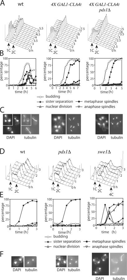
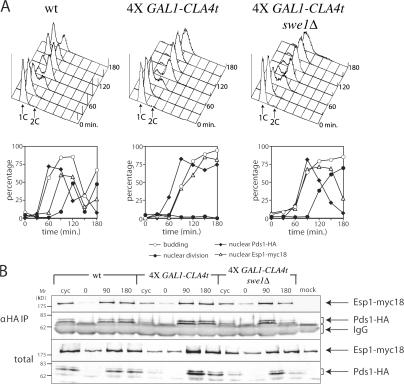
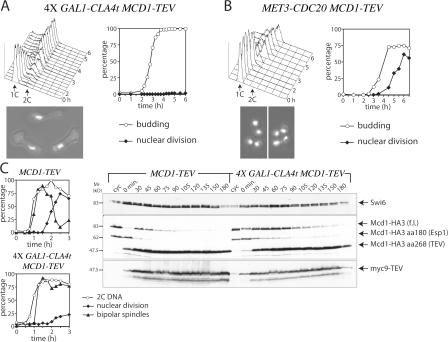
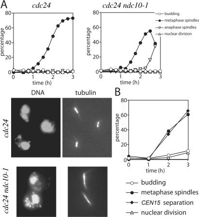
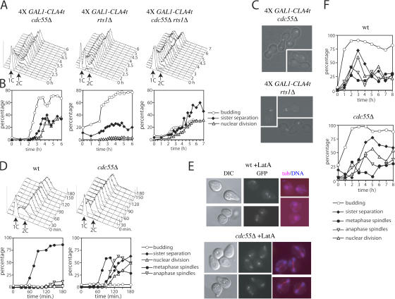
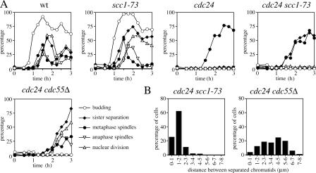
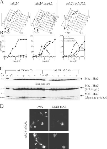
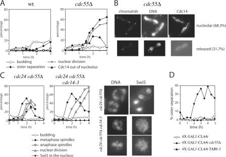
Faithful chromosome transmission requires establishment of sister chromatid cohesion during S phase, followed by its removal at anaphase onset. Sister chromatids are tethered together by cohesin, which is displaced from chromosomes through cleavage of its Mcd1 subunit by the separase protease. Separase is in turn inhibited, up to this moment, by securin. Budding yeast cells respond to morphogenetic defects by a transient arrest in G2 with high securin levels and unseparated chromatids. We show that neither securin elimination nor forced cohesin cleavage is sufficient for anaphase in these conditions, suggesting that other factors contribute to cohesion maintainance in G2. We find that the protein phosphatase PP2A bound to its regulatory subunit Cdc55 plays a key role in this process, uncovering a new function for PP2A(Cdc55) in controlling a noncanonical pathway of chromatid cohesion removal.


Figures

Figure 1.
Figure 1.
G2 arrest by the morphogenesis checkpoint does not depend on securin. (A–C) Strains with the indicated genotypes (ySP3575, ySP3435, and ySP3436) were grown at 25°C in YEPR. Elutriated G1 unbudded cells were released at 25°C in the presence of galactose (time 0), followed by FACS analysis of DNA contents (A) and determination of kinetics of budding, sister chromatid separation, spindle formation/elongation, and nuclear division (B). Micrographs (C) were taken at 240 min (wt), 420 min (4X GAL1-CLA4t), and 300 min (4X GAL1-CLA4t pds1Δ). (D–F) wt (ySP601), pds1Δ (ySP2894), and swe1Δ (ySP3887) cells were grown in YEPD at 23°C, arrested in G1 by α-factor, and released in the presence of Lat-A, followed by FACS analysis of DNA contents (D) and determination of kinetics of budding, sister chromatid separation, spindle formation/elongation, and nuclear division (E). (F) Micrographs represent cells at 90 min.
Figure 2.
Figure 2.
Binding to securin and nuclear accumulation of separase are not affected by the morphogenesis checkpoint. Strains with the indicated genotypes (ySP1735, ySP4690, and ySP4692) were grown in YEPR at 25°C, arrested by α-factor, and released in YEPRG (time 0). (A) At the indicated times, cells were analyzed for DNA contents (histograms), budding, nuclear division, and Pds1 and Esp1 nuclear accumulation (graphs). Pds1 and Esp1 localization was scored on fixed cells after in situ immunofluorescence. (B) Protein extracts from cells harvested at the indicated times were analyzed by immunoblotting with anti-HA (Pds1) or anti-myc (Esp1) antibodies either directly (total) or after Pds1 immunoprecipitation with anti-HA antibodies (αHA IP).
Figure 3.
Figure 3.
Mcd1 cleavage is not sufficient for nuclear division upon CLA4t overexpression. (A) GAL1-CLA4t cells expressing Mcd1-TEV and GAL1-TEV (ySP5871) were grown in YEPR at 25°C. Elutriated small G1 cells were released in YEPRG at 25°C (time 0). At the indicated time points, cell samples were analyzed for DNA contents (top left), budding, and nuclear division (top right). Micrographs represent cells at the end of the experiment. (B) MET3-CDC20 MCD1-TEV GAL1-TEV cells (ySP5870) were grown in raffinose medium lacking methionine. Elutriated G1 cells were released in YEPRG containing 2 mM methionine (time 0). Cell samples were analyzed as in A. (C) MCD1-TEV (ySP3448) and GAL1-CLA4t MCD1-TEV (ySP5871) cells were grown in YEPR at 25°C, arrested in G1 by α-factor, and released in YEPRG at 25°C at time 0. Cells were collected at the indicated times for Western blot analysis with anti-HA (Mcd1) and anti-myc (TEV) antibodies (left), FACS analysis of DNA contents (not depicted), and kinetics of nuclear division and bipolar spindle formation (right). Swi6 was used as loading control.
Figure 4.
Figure 4.
Spindle dynamics is not affected by the morphogenesis checkpoint. (A) cdc24 (ySP305) and cdc24 ndc10-1 (ySP6207) cell cultures were arrested in G1 by α-factor and released at 37°C (time 0). Cells were analyzed at the indicated times for DNA contents (not depicted), budding, spindle formation/elongation, and nuclear division (graphs). Micrographs represent cells at 150 min after release. (B) Wild-type cells with tagged CEN15 (ySP1717) were arrested in G1 by α-factor and released in the presence of Lat-B (time 0). Cells were analyzed at 1-h intervals for DNA contents (not depicted), CEN15 separation, tubulin immunostaining, and nuclear division.
Figure 5.
Figure 5.
PP2ACdc55 prevents sister chromatid separation upon activation of the morphogenesis checkpoint. (A–C) Strains with the indicated genotypes (ySP5115, ySP5112, and ySP5165) were grown at 30°C in YEPR. Elutriated G1 cells were released in YEPRG at 25°C at time 0. Cell samples were analyzed at the indicated times for DNA contents (A), budding, sister chromatid separation, and nuclear division (B). (C) Micrographs represent sister chromatid separation at 285 min (4X GAL1-CLA4t cdc55Δ) and 300 min (4X GAL1-CLA4t rts1Δ). (D) Wild-type (wt; ySP3575) and cdc55Δ (ySP5068) cells were grown in YEPD at 30°C, arrested in G1 with α-factor, and released in the presence of Lat-A (time 0). Cells were analyzed at the indicated times for DNA contents (histograms), budding, sister separation, nuclear division, and spindle formation/elongation (graphs). (E) Micrographs represent pericentromeric chromosomal sequences (GFP) and merged pictures of tubulin and DNA staining (tub/DNA) at 150 min. DIC, differential interference contrast. (F) Wild-type (ySP3575) and cdc55Δ (ySP5068) cells were arrested in G1 by α-factor and released at 16°C. Cells were analyzed at the indicated times for budding, sister chromatid separation, spindle formation/elongation, and nuclear division.
Figure 6.
Figure 6.
Inactivation of PP2ACdc55, but not of cohesin, allows nuclear division in the presence of morphogenetic defects. Strains with the indicated genotypes (ySP601, ySP818, ySP6236, ySP6241, and ySP6214) were arrested in G1 by α-factor at 25°C and released at 37°C (time 0). Cells were analyzed at the indicated times for DNA contents (not depicted), budding, sister chromatid separation, spindle formation/elongation, and nuclear division (A). Distances between separated chromatids were measured at time point 150 min (n = 180) with MetaMorph software.
Figure 7.
Figure 7.
Lack of Cdc55 upon Cla4t overproduction allows anaphase in the absence of Mcd1 cleavage. Strains with the indicated genotypes (ySP6249, ySP6250, and ySP6463) growing at 25°C were arrested in G1 by α-factor and released at 37°C (time 0). At the indicated times, cell samples were collected for FACS analysis (A); kinetics of budding, sister chromatid separation, and spindle formation/elongation (B); Western blot analysis of TCA protein extracts (C); and chromosome spreads (D). Total extracts were immunoblotted with anti-HA antibodies to detect Mcd1-HA3 (C). Chromosome spreads (D) were immunostained with anti-HA antibodies (Mcd1-HA3) and mounted with DAPI to stain DNA. Arrows indicate anaphase nuclei.
Figure 8.
Figure 8.
Cdc14 is not required for nuclear division of cdc55Δ cells with morphogenetic defects. (A) Wild-type (wt; ySP3575) and cdc55Δ (ySP5068) cells were arrested in G1 by α-factor at 30°C and released at 25°C in the presence of Lat-B (time 0). At the indicated times, cells were collected to determine the kinetics of budding, sister chromatid separation, nuclear division, and Cdc14 nucleolar export after in situ immunofluorescence. Micrographs (B) show examples of cdc55Δ cells at 150 min after release. (C) cdc24 cdc55Δ (ySP6503) and cdc24 cdc55Δ cdc14-3 (ySP6499) cells expressing myc-tagged Swi5 were arrested in G1 by α-factor at 25°C and released at 37°C (time 0). Cells were analyzed at the indicated times for budding, spindle formation/elongation, nuclear division, and Swi5 nuclear import. Pictures were taken at 150 min after release. (D) Strains with the indicated genotypes (ySP5704, ySP5710, and ySP5893) were grown in YEPR at 30°C, arrested in G1 by α-factor, and released in YEPRG at 25°C (time 0), followed by scoring GFP dots to determine the kinetics of sister chromatid separation.
Figure 9.
Figure 9.
Effects of Cdc5, condensin, and topoisomerase II inactivation on anaphase onset in the absence of Cdc55. (A) Strains with the indicated genotypes (W303, ySP305, ySP6130, ySP6146, ySP6121, and ySP6105) were grown in YEPD at 26°C, arrested in G1 with α-factor, and released at 37°C (time 0). At different times, cells were collected to determine the kinetics of budding and anaphase after nuclear staining with propidium iodide. (B) Serial dilutions of strains with the indicated genotypes (W303, ySP5737, ySP5929, and ySP6066) were spotted on YEPD plates and incubated at 25°C for 2 d and at 16°C for 6 d. (C) Wild-type (wt), top2-4, and cdc24 cells carrying a minichromosome (YCp50) were arrested in G1 with α-factor and released for 3 h at 37°C in the presence of nocodazole. Topological forms of plasmids isolated from these cells were examined by Southern blot after electrophoresis of total DNA in the presence of ethidium bromide using the 1.7-kb AmpR PvuI–BglII fragment of YCp50 as a probe. Purified YCp50 DNA linearized with HindIII and uncut were run in parallel to define the mobility of the various conformational forms.
Figure 10.
Figure 10.
CDC55 overexpression delays anaphase independently of securin. Wild-type (wt; ySP3575), GAL1-CDC55 (ySP5690), and GAL1-CDC55 pds1Δ (ySP5752) strains carrying the tetO/tetR-GFP construct to detect pericentromeric sequences at chromosome V were grown in YEPR at 25°C, arrested in G1 by α-factor, and released in YEPRG at 25°C (time 0). Cells were analyzed at different times for DNA contents (histograms), budding, sister chromatid separation, spindle formation/elongation, and nuclear division (graphs). Pictures show merged micrographs of tubulin (red) and DNA (blue) at 150 min.

Similar articles

Cited by

References

    1. Agarwal, R., and O. Cohen-Fix. 2002. Mitotic regulation: the fine tuning of separase activity. Cell Cycle. 1:255–257. - PubMed
    1. Alexandru, G., F. Uhlmann, K. Mechtler, M. Poupart, and K. Nasmyth. 2001. Phosphorylation of the cohesin subunit Scc1 by Polo/Cdc5 kinase regulates sister chromatid separation in yeast. Cell. 105:459–472. - PubMed
    1. Bachant, J., A. Alcasabas, Y. Blat, N. Kleckner, and S.J. Elledge. 2002. The SUMO-1 isopeptidase Smt4 is linked to centromeric cohesion through SUMO-1 modification of DNA topoisomerase II. Mol. Cell. 9:1169–1182. - PubMed
    1. Bhalla, N., S. Biggins, and A.W. Murray. 2002. Mutation of YCS4, a budding yeast condensin subunit, affects mitotic and nonmitotic chromosome behavior. Mol. Biol. Cell. 13:632–645. - PMC - PubMed
    1. Charles, J.F., S.L. Jaspersen, R.L. Tinker-Kulberg, L. Hwang, A. Szidon, and D.O. Morgan. 1998. The Polo-related kinase Cdc5 activates and is destroyed by the mitotic cyclin destruction machinery in S. cerevisiae. Curr. Biol. 8:497–507. - PubMed

Publication types

MeSH terms