. 2007 May 21;177(4):613-24.
doi: 10.1083/jcb.200611063. Epub 2007 May 14.

Ubiquitination of serine, threonine, or lysine residues on the cytoplasmic tail can induce ERAD of MHC-I by viral E3 ligase mK3

Affiliations

Ubiquitination of serine, threonine, or lysine residues on the cytoplasmic tail can induce ERAD of MHC-I by viral E3 ligase mK3

Xiaoli Wang et al. J Cell Biol. .

Abstract

The mechanism by which substrates for endoplasmic reticulum-associated degradation are retrotranslocated to the cytosol remains largely unknown, although ubiquitination is known to play a key role. The mouse gamma-herpesvirus protein mK3 is a viral RING-CH-type E3 ligase that specifically targets nascent major histocompatibility complex I heavy chain (HC) for degradation, thus blocking the immune detection of virus-infected cells. To address the question of how HC is retrotranslocated and what role mK3 ligase plays in this action, we investigated ubiquitin conjugation sites on HC using mutagenesis and biochemistry approaches. In total, our data demonstrate that mK3-mediated ubiquitination can occur via serine, threonine, or lysine residues on the HC tail, each of which is sufficient to induce the rapid degradation of HC. Given that mK3 has numerous cellular and viral homologues, it will be of considerable interest to determine the pervasiveness of this novel mechanism of ubiquitination.


Figures

Figure 1.
Figure 1.
The K residues of MHC class I HC are dispensable for mK3-mediated HC down-regulation. (A) The structure of the ectodomain of Ld was generated using published coordinates of the Ld–p29 complex (Protein Data Bank 1LD9). There are a total of 12 K residues on the Ld molecule: the three on the cytoplasmic tail are K308, K316, and K337, and the nine on the ectodomain are shown in the structure. Of the nine, six conserved Ks are underlined, and the other three (K31, K173, and K196) are unique to Ld. (B) WT3 cells expressing wild-type (wt) or K-less Ld were cotransduced with one of the following constructs: pMIG only (vector), pMIG.mK3 (mK3), or pMIG.mK3 C48G, C51G (an mK3 RING-CH domain mutant named mK3 RING mut). Flow cytometric analysis of surface Ld expression (detected by 30-5-7 staining) versus GFP fluorescence is shown. The number in each plot represents the ratio of Ld staining intensities between GFP+ and GFP populations. (C) Cells used in B were enriched by sorting the GFP+ fraction of the transduced cell line. These enriched cells were incubated for 24 h with 125 U/ml IFN-γ, pulse labeled with [35S]Cys/Met, and chased for the indicated times with unlabeled Cys/Met. Ld precipitates were resolved by SDS-PAGE and visualized by autoradiography. Ld and β2m bands are indicated (left). In the right panel, relative band intensities from the gels are plotted as a percentage of the intensity at time zero for each cell line.
Figure 2.
Figure 2.
Down-regulation of completely K-less Ld is ubiquitination-proteasome dependent. (A) Ld HCs were immunoprecipitated with mAbs (30-5-7 and 64-3-7) from NP-40 lysates of the cell lines used in Fig. 1 B. Precipitates with or without endo H treatment were separated by SDS-PAGE and blotted for Ub and Ld HC. In the right panel, the β-actin blot was included as a loading control. (B) After incubation for 2 h with 5 μM/ml of the proteasome inhibitor (PI) clasto-lactacystin β-lactone, cells expressing wt Ld or K-less Ld (±mK3) were harvested and NP-40 lysed. Ld HCs were precipitated, treated, or mock treated with endo H and were blotted with the indicated antibodies. (C) Lysates from B were blotted for mK3 (rabbit anti-mK3). The β-actin blot was included to control for equal loading.
Figure 3.
Figure 3.
Ubiquitination and degradation of Ub/Ld fusion proteins in the presence of mK3. (A) After incubation for 24 h with 125 U/ml IFN-γ, WT3 cells expressing Ub wt/Ld K-less or Ub K-less/Ld K-less (±mK3) were pulse labeled with [35S]Cys/Met for 15 min and chased for the indicated times. Ub/Ld fusion molecules were precipitated by anti-Ld mAbs. Precipitates then were resolved by SDS-PAGE and visualized by autoradiography (left). Relative band intensities from these gels were plotted as a percentage of the intensity at time zero for each line (right). (B) Cells used in A were Dounce homogenized. The homogenate was then subjected to serial centrifugations from 1,000 to 100,000 g. After digestion (or mock digestion) with 10 μg/ml proteinase K for 20 min on ice, Ub/Ld molecules were precipitated from NP-40 lysates of 100,000 g pellet (P) or supernatant (S) and analyzed by immunoblotting using anti-Ub mAb or rabbit anti-Ld cytoplasmic tail (cyt tail) antibodies. Polyubiquitinated forms are indicated by asterisks.
Figure 4.
Figure 4.
Polyubiquitinated Ld HCs were no longer detectable when the cytoplasmic tail of HC was removed. (A) Similar procedures as described in Fig. 3 B were used for analysis of the cells expressing wt Ld (±mK3). Precipitates of Ld HCs were immunoblotted by anti-Ub mAb, anti-Ld lumenal mAbs (64-3-7), and anti-Ld cytoplasmic tail antibodies. Polyubiquitinated forms are indicated by asterisks. (B) Ld HCs were precipitated from NP-40 lysates of the cells coexpressing mK3 and wt Ld or mK3 and Ld TMB (wt Ld with a K308R mutation and an engineered TMB cleavage site in its cytoplasmic tail; the tail sequence of this molecule is VMRRRRNTLVPRGGKGGDYALAPGSQSSEMSLRDCKA, with the TMB cleavage site in bold). After incubation with TMB at room temperature for 3 h and then at 4°C overnight, the precipitates were analyzed by immunoblotting using the indicated antibodies.
Figure 5.
Figure 5.
The C residue in the tail of Ld HC is not required for its ubiquitination and rapid degradation mediated by mK3. (A) Sequence alignment of the cytoplasmic tails of wt Ld and a tail KC-less mutant is shown with wt residues in bold and substituted residues in red. (B) Polyubiquitination of Ld HCs in the cells expressing wt Ld or tail KC-less Ld (±mK3) was examined by immunoprecipitation of Ld HCs from NP-40 lysates (±endo H treatment) and immunoblotting for Ub. (C) After incubation for 24 h with 125 U/ml IFN-γ, cells used in B were pulse labeled with [35S]Cys/Met for 15 min and chased for the indicated times. Ld HCs were precipitated, resolved, and visualized as described in Fig. 1 C. Relative band intensities from the gels are plotted as a percentage of the intensity at time zero for each line and are shown beside the gel photos. (D) After incubation with 60 μM MG132 for 2 h, wt Ld HCs or K-less Ld HCs were precipitated by anti-Ld mAbs and eluted in the presence or absence of 5% 2ME at pH 8.0. Precipitates then were analyzed by immunoblotting for Ub and Ld HC. Ig bands are indicated by arrows; nonreduced or reduced (non-rd or rd) Ld HC bands and poly-Ub forms (Ubn) are also indicated.
Figure 6.
Figure 6.
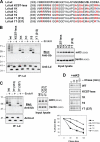
Ubiquitination of Ld by mK3 can occur on either S, T, or K residues on the tail of Ld. (A) Nomenclature and sequence alignment of the cytoplasmic tails of Ld tail mutants are shown with original wt residues in bold, substituted residues in red, and added back residues in blue. (B) WT3 cells stably coexpressing mK3 and one of the Ld tail mutants were NP-40 lysed. After precipitation of Ld, precipitates were immunoblotted for Ub and Ld (left). In the right panel, the β-actin and mK3 blots were included to show that a similar amount of input lysate of each line was used for immunoprecipitation and a similar amount of mK3 was expressed in each line. (C) Cells coexpressing mK3 and one of the Ld tail mutants 1K, 1T(313), or 1T(337) were lysed, immunoprecipitated, and blotted as described in B. (D) After incubation for 24 h with 125 U/ml IFN-γ, cells used in B and C were pulse labeled with [35S]Cys/Met for 15 min and chased for the indicated times. Ld HCs were precipitated, resolved, and visualized as described in Fig. 1 C. Relative band intensities from the gels are plotted as a percentage of the intensity at time zero for each line and are shown beneath the gel photos.
Figure 7.
Figure 7.
Evidence for the direct conjugation of Ub to an S residue of the Ld tail mediated by mK3. (A) Ld HCs were precipitated by anti-Ld mAbs 30-5-7 and 64-3-7 from NP-40 lysates of the cells expressing Ld tail 1K or Ld tail 1S mutant (±mK3). After incubation in 0.5% SDS and 10 mM DTT at room temperature for 10 min and boiling for 10 min, part of the elutes were reprecipitated by the conformation-independent anti-Ld mAb 64-3-7. Note that for this experiment, similar amounts of Ld were loaded for each sample. The first and second precipitates were immunoblotted for Ub, Ld, and β2m. Ub-conjugated Ld forms (Ubn-Ld) and unconjugated Ld HCs are indicated. Nonspecific staining of antibody heavy chain (Ig HC) is also indicated. (B) After incubation with 30 μM MG132 for 3 h, the cells stably expressing Ld tail KCST-less, Ld tail 1K, or Ld tail 1S (+mK3) were lysed with NP-40. After precipitation of Ld, the precipitates were incubated with or without (−/+) 1 M sodium hydroxylamine, pH 9, for 4 h at 37°C (top) or with or without (−/+) 0.1 M NaOH for 2 h at 37°C (bottom). Then, the precipitates were subjected to SDS-PAGE and immunoblotting for Ub and Ld. Ub-conjugated Ld forms (Ub2-Ld or Ub3-Ld) and unconjugated Ld HCs are indicated. Because NaOH-treated samples were reduced by 2.5% 2ME before loading for SDS-PAGE, unconjugated and conjugated Ld HCs migrate slower than nonreduced sodium hydroxylamine–treated samples, and a disassociated Ig HC band can be seen in the blot.

Similar articles

Cited by

References

    1. Baker, R.T. 1996. Protein expression using ubiquitin fusion and cleavage. Curr. Opin. Biotechnol. 7:541–546. - PubMed
    1. Barel, M.T., N. Pizzato, D. van Leeuwen, P.L. Bouteiller, E.J. Wiertz, and F. Lenfant. 2003. Amino acid composition of alpha1/alpha2 domains and cytoplasmic tail of MHC class I molecules determine their susceptibility to human cytomegalovirus US11-mediated down-regulation. Eur. J. Immunol. 33:1707–1716. - PubMed
    1. Bartee, E., M. Mansouri, B.T. Hovey Nerenberg, K. Gouveia, and K. Fruh. 2004. Downregulation of major histocompatibility complex class I by human ubiquitin ligases related to viral immune evasion proteins. J. Virol. 78:1109–1120. - PMC - PubMed
    1. Bhamidipati, A., V. Denic, E.M. Quan, and J.S. Weissman. 2005. Exploration of the topological requirements of ERAD identifies Yos9p as a lectin sensor of misfolded glycoproteins in the ER lumen. Mol. Cell. 19:741–751. - PubMed
    1. Biederer, T., C. Volkwein, and T. Sommer. 1997. Role of Cue1p in ubiquitination and degradation at the ER surface. Science. 278:1806–1809. - PubMed

Publication types

MeSH terms