

Role of Peyer's patches in the induction of Helicobacter pylori-induced gastritis
- PMID: 17502608
- PMCID: PMC1885612
- DOI: 10.1073/pnas.0609014104
Role of Peyer's patches in the induction of Helicobacter pylori-induced gastritis
Abstract
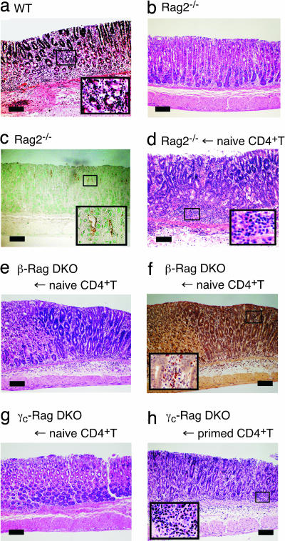
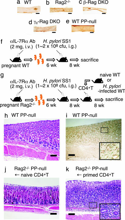
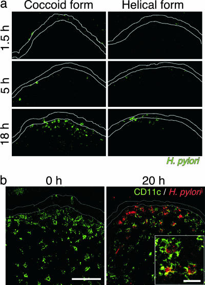
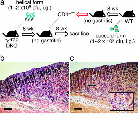
Helicobacter pylori is a Gram-negative spiral bacterium that causes gastritis and peptic ulcer and has been implicated in the pathogenesis of gastric adenocarcinoma and mucosa-associated lymphoid tissue lymphoma. Although Th1 immunity is involved in gastritis and the accumulation of H. pylori-specific CD4(+) T cells in the H. pylori-infected gastric mucosa in human patients, how T cells are primed with H. pylori antigens is unknown because no apparent lymphoid tissues are present in the stomach. We demonstrate here that Peyer's patches (PPs) in the small intestine play critical roles in H. pylori-induced gastritis; no gastritis is induced in H. pylori-infected mice lacking PPs. We also observed that the coccoid form of H. pylori is phagocytosed by dendritic cells in PPs. We propose that H. pylori converts to the coccoid form in the anaerobic small intestine and stimulates the host immune system through PPs.
Conflict of interest statement
The authors declare no conflict of interest.
Figures
Comment in
-
In"sight" into immune blindness: Helicobacter pylori-induced gastritis requires T cells primed in the small intestine Peyer's patches.Gastroenterology. 2007 Sep;133(3):1044-5. doi: 10.1053/j.gastro.2007.07.044. Gastroenterology. 2007. PMID: 17854610 No abstract available.
Similar articles
-
Essential role of Peyer's patches in the development of Helicobacter-induced gastritis.Int Immunol. 2007 Apr;19(4):435-46. doi: 10.1093/intimm/dxm008. Epub 2007 Feb 20. Int Immunol. 2007. PMID: 17314082
-
Small intestine Peyer's patches are major induction sites of the Helicobacter-induced host immune responses.Gastroenterology. 2008 Feb;134(2):642-3. doi: 10.1053/j.gastro.2007.12.007. Gastroenterology. 2008. PMID: 18242237 No abstract available.
-
Helicobacter heilmannii can induce gastric lymphoid follicles in mice via a Peyer's patch-independent pathway.FEMS Immunol Med Microbiol. 2010 Nov;60(2):156-64. doi: 10.1111/j.1574-695X.2010.00731.x. Epub 2010 Sep 16. FEMS Immunol Med Microbiol. 2010. PMID: 20846360
-
Helicobacter pylori immunology and vaccines.Helicobacter. 2008 Oct;13 Suppl 1:18-22. doi: 10.1111/j.1523-5378.2008.00636.x. Helicobacter. 2008. PMID: 18783517 Review.
-
The design of vaccines against Helicobacter pylori and their development.Annu Rev Immunol. 2001;19:523-63. doi: 10.1146/annurev.immunol.19.1.523. Annu Rev Immunol. 2001. PMID: 11244046 Review.
Cited by
-
Effects of Helicobacter pylori infection on intestinal microbiota, immunity and colorectal cancer risk.Front Cell Infect Microbiol. 2024 Jan 26;14:1339750. doi: 10.3389/fcimb.2024.1339750. eCollection 2024. Front Cell Infect Microbiol. 2024. PMID: 38343887 Free PMC article. Review.
-
DC-derived IL-18 drives Treg differentiation, murine Helicobacter pylori-specific immune tolerance, and asthma protection.J Clin Invest. 2012 Mar;122(3):1082-96. doi: 10.1172/JCI61029. Epub 2012 Feb 6. J Clin Invest. 2012. PMID: 22307326 Free PMC article.
-
IL-23 Contributes to Control of Chronic Helicobacter Pylori Infection and the Development of T Helper Responses in a Mouse Model.Front Immunol. 2012 Mar 26;3:56. doi: 10.3389/fimmu.2012.00056. eCollection 2012. Front Immunol. 2012. PMID: 22566937 Free PMC article.
-
Indigenous opportunistic bacteria inhabit mammalian gut-associated lymphoid tissues and share a mucosal antibody-mediated symbiosis.Proc Natl Acad Sci U S A. 2010 Apr 20;107(16):7419-24. doi: 10.1073/pnas.1001061107. Epub 2010 Apr 1. Proc Natl Acad Sci U S A. 2010. PMID: 20360558 Free PMC article.
-
Progress in elucidating the relationship between Helicobacter pylori infection and intestinal diseases.World J Gastroenterol. 2021 Dec 21;27(47):8040-8046. doi: 10.3748/wjg.v27.i47.8040. World J Gastroenterol. 2021. PMID: 35068852 Free PMC article.
References
-
- Covacci A, Telford JL, Del Giudice G, Parsonnet J, Rappuoli R. Science. 1999;284:1328–1333. - PubMed
-
- Ernst PB, Gold BD. Annu Rev Microbiol. 2000;54:615–640. - PubMed
-
- Uemura N, Okamoto S, Yamamoto S, Matsumura N, Yamaguchi S, Yamakido M, Taniyama K, Sasaki N, Schlemper RJ. N Engl J Med. 2001;345:784–789. - PubMed
-
- Baldari CT, Lanzavecchia A, Telford JL. Trends Immunol. 2005;26:199–207. - PubMed
-
- Cover TL, Blanke SR. Nat Rev Microbiol. 2005;3:320–332. - PubMed
Publication types
MeSH terms
Substances
LinkOut - more resources
Full Text Sources
Other Literature Sources
Medical
Research Materials
