. 2007 May 22;104(21):8755-60.
doi: 10.1073/pnas.0702290104. Epub 2007 May 14.

Trans-SNARE complex assembly and yeast vacuole membrane fusion

Affiliations

Trans-SNARE complex assembly and yeast vacuole membrane fusion

Kevin M Collins et al. Proc Natl Acad Sci U S A. .

Abstract

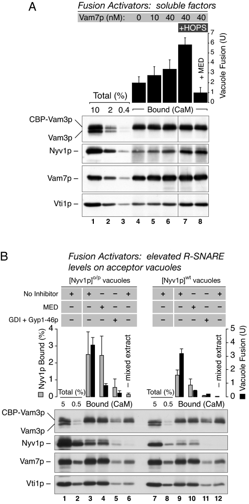
cis-SNARE complexes (anchored in one membrane) are disassembled by Sec17p (alpha-SNAP) and Sec18p (NSF), permitting the unpaired SNAREs to assemble in trans. We now report a direct assay of trans-SNARE complex formation during yeast vacuole docking. SNARE complex assembly and fusion is promoted by high concentrations of the SNARE Vam7p or Nyv1p or by addition of HOPS (homotypic fusion and vacuole protein sorting), a Ypt7p (Rab)-effector complex with a Sec1/Munc18-family subunit. Inhibitors that target Ypt7p, HOPS, or key regulatory lipids prevent trans-SNARE complex assembly and ensuing fusion. Strikingly, the lipid ligand MED (myristoylated alanine-rich C kinase substrate effector domain) or elevated concentrations of Sec17p, which can displace HOPS from SNARE complexes, permit full trans-SNARE pairing but block fusion. These findings suggest that efficient fusion requires trans-SNARE complex associations with factors such as HOPS and subsequent regulated lipid rearrangements.


Conflict of interest statement

The authors declare no conflict of interest.

Figures

Fig. 1.
Fig. 1.
Vam3p bearing an internal epitope tag is functional in vivo. (A) Vam3p domain structure. A CBP was inserted between the helices ABC N-terminal domain and the SNARE domain. The CBP was inserted N-terminal to the dileucine sorting motif. (BF) Vacuole morphology in yeast cells expressing wild-type Vam3p or CBP-Vam3p. Overnight cultures of BJ3505 yeast cells and derivatives were stained in YPD with FM 4-64 (5 μM). Cells were concentrated by centrifugation, applied to glass slides, and overlaid with a coverslip. Micrographs of stained vacuoles were acquired as described (6). (G) Protein composition of isolated yeast vacuoles. Vacuoles from the indicated strains were heated in sample buffer. Proteins were separated by SDS/PAGE (5 μg per lane), transferred to nitrocellulose, and incubated with the indicated antibodies. Because vacuoles prepared from DKY6281 have active proteases, some protein degradation (e.g., Snc1/2p and Vps41p) occurred during isolation.
Fig. 2.
Fig. 2.
CBP-Vam3p promotes vacuole fusion and SNARE complex assembly in vitro. (A) CBP-Vam3p, as the sole source of Vam3p, can mediate vacuole fusion. Vacuoles were isolated from the indicated strains (pep4Δ is BJ3505 and pho8Δ is DKY6281) expressing the indicated SNAREs and assayed under standard fusion conditions. Reactions were incubated at 27°C (or held on ice) for 90 min. Where indicated, GDI (1 μM), Sec18p (1.5 nM), or Vam7p (40 nM) were added from the start. Bars represent the average fusion activity of two independent experiments in which reactions bearing wild-type Vam3p and no fusion activator or inhibitor (middle gray bar) was set to 100%. Error bars indicate the range of the values in the two experiments. The asterisk highlights that combination of yeast vacuoles used for subsequent trans-SNARE interactions in this study. (B) Assay of SNARE complex assembly between CBP-Vam3p and Nyv1p. Vacuoles bearing CBP-Vam3p and deleted for Nyv1p were mixed with vacuoles from DKY6281. Reactions were incubated at 27°C for the indicated times after which vacuole fusion (black bars) and the associations of CBP-Vam3p with Nyv1p from the acceptor vacuoles (gray bars) and other SNAREs was measured by affinity purification from detergent extracts using immobilized calmodulin on agarose beads (see Materials and Methods). Two detergent extracts were also prepared from reactions containing a single vacuole type, a CBP-Vam3p nyv1Δ vacuole reaction, or a DKY6281 vacuole reaction. These two extracts were mixed and the CBP-Vam3p interactions with Nyv1p that occur in solution were examined (lane 10). The experiment shown was performed in duplicate and is representative of three independent trials. Bars indicate the average fusion and Nyv1p associations as determined by densitometry of the duplicates, and error bars represent the range of values between them.
Fig. 3.
Fig. 3.
Control of trans-SNARE complex assembly. (A) Testing cis-SNARE disassembly inhibitors and their bypass by Vam7p. Fusion reactions containing trans-SNARE tester vacuoles were incubated with buffer alone (lanes 4 and 8), antibodies to Sec17p (lanes 5 and 9) or Sec18p (lanes 6 and 10), or excess Sec17p (750 nM; lanes 7 and 11). Vam7p was then added where indicated (lanes 8–11). Reactions were incubated (27°C, 45 min) and fusion (black bars) and CBP-Vam3p associations with Nyv1p (gray bars) and other SNAREs were assayed. Data are representative of three independent experiments. Bars represent the average fusion and Nyv1p associations found in three (without Vam7p) or four experiments (with Vam7p). Error bars indicate the standard deviation of the values obtained. (B) Role of Ypt7p and HOPS. Fusion reactions with 40 nM Vam7p were incubated with buffer alone (lanes 4 and 5), GDI (lane 6), Gyp1-46p (lane 7), GDI and Gyp1-46p (lane 8), antibodies to Ypt7p (lane 9), antibodies to Ypt7p premixed with cognate peptide (lane 10), or antibodies to Vps33p (lane 11). Reactions were incubated (27°C, 60 min), and fusion (Top) and CBP-Vam3p association with other SNAREs assayed. Fusion values are the average of three independent experiments and error bars represent the standard deviation. SNARE associations are representative of three independent experiments.
Fig. 4.
Fig. 4.
Regulatory lipid requirements during trans-SNARE complex assembly. Fusion reactions were incubated with Vam7p with no added inhibitors (lane 1) or with the indicated inhibitors (lanes 2–6). After incubation (45 min, 27°C), reactions were placed on ice and fusion (black bars) and CBP-Vam3p associations with Nyv1p (gray bars) and other SNAREs were assayed. Data are representative of four experiments whose results were averaged; error bars represent the standard deviation of the values obtained after densitometric analysis of Nyv1p associations.
Fig. 5.
Fig. 5.
Promotion of trans-SNARE complex assembly. (A) Added Vam7p and HOPS. Fusion reactions contained added Vam7p. Purified HOPS complex (5 μg/ml) was added after Vam7p addition (lanes 7 and 8), and MED (lane 8) was added to one HOPS + Vam7p reaction. Vacuole fusion and CBP-Vam3p associations were assayed after incubation (45 min, 27°C). Data are representative of three independent experiments. Bars represent the average fusion data from the three trials, and error bars represent their standard deviation. (B) Elevated Nyv1p on acceptor vacuoles. Fusion reactions were initiated with vacuoles from BJ3505 CBP-Vam3p nyv1Δ and a DKY6281 strain which either overproduced Nyv1p from the ADH1 promoter element ([Nyv1p]o/p, lanes 1–6; Y. Jun and WTW, unpublished data) or bore wild type levels of Nyv1p ([Nyv1p]wt, lanes 7–12). After incubation (45 min, 27°C), reactions were placed on ice, and fusion (black bars) and associations of CBP-Vam3p with Nyv1p (gray bars) and other SNAREs were assayed. Vacuole detergent extracts from reactions containing a single type of vacuole (CBP-Vam3p nyv1Δ, Nyv1po/p, or Nyv1pwt) were also prepared as in Fig. 2B. These reaction extracts were mixed to determine the extent of Nyv1p associations that occurred in the extract (lanes 6 and 12). Three independent experiments were averaged, and error bars represent the standard deviation of the fusion values and the Nyv1p associations as determined by densitometry.

Similar articles

Cited by

References

    1. Jahn R, Scheller RH. Nat Rev Mol Cell Biol. 2006;7:631–643. - PubMed
    1. Sutton RB, Fasshauer D, Jahn R, Brunger AT. Nature. 1998;395:347–353. - PubMed
    1. Fasshauer D, Sutton RB, Brunger AT, Jahn R. Proc Natl Acad Sci USA. 1998;95:15781–15786. - PMC - PubMed
    1. Ungermann C, Sato K, Wickner W. Nature. 1998;396:543–548. - PubMed
    1. Carr CM, Grote E, Munson M, Hughson FM, Novick PJ. J Cell Biol. 1999;146:333–344. - PMC - PubMed

Publication types

MeSH terms