. 2007 May 22;104(21):8833-8.
doi: 10.1073/pnas.0701666104. Epub 2007 May 14.

Exposing the core promoter is sufficient to activate transcription and alter coactivator requirement at RNR3

Affiliations

Exposing the core promoter is sufficient to activate transcription and alter coactivator requirement at RNR3

Hesheng Zhang et al. Proc Natl Acad Sci U S A. .

Abstract

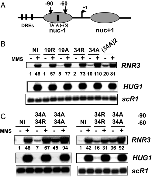
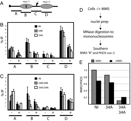
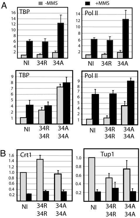
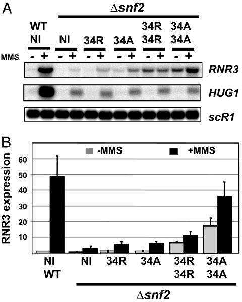
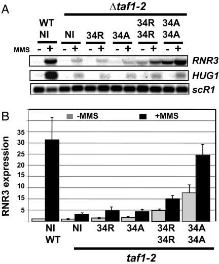
Chromatin is a formidable barrier to transcription. Nucleosome density is lowest over the regulatory regions of active genes, and many repressed genes have a tightly positioned nucleosome over their core promoter. However, it has not been shown that nucleosome positioning is sufficient for repression or whether disrupting a core promoter nucleosome specifically can activate gene expression in the absence of activating signals. Here we show that disrupting the nucleosome over the core promoter of RNR3 is sufficient to drive preinitiation complex assembly and activate transcription in the absence of activating signals. Remodeling of chromatin over the RNR3 promoter requires the recruitment of the SWI/SNF complex by the general transcription factor TFIID. We found that disrupting the nucleosome over the RNR3 core promoter relieves its dependence on TFIID and SWI/SNF, indicating a functional link between these two complexes. These results suggest that the specific function of TAF(II)s is to direct the chromatin remodeling step through SWI/SNF recruitment, and not core promoter selectivity. Our results indicate that nucleosome placement plays a dominant role in repression and that the ability of the core promoter to position a nucleosome is a major determinant in TAF(II) dependency of genes in vivo.


Conflict of interest statement

The authors declare no conflict of interest.

Figures

Fig. 1.
Fig. 1.
Insertion of PNTs within RNR3. (A) A schematic of RNR3 and its chromatin structure based on Li and Reese (13). (B) Random DNA sequence (R) or poly dA:dT (A) of differing lengths were inserted upstream (−90) of the TATA box, which is located at −75 relative to the start site of transcription (black box). Shown are Northern blots of RNR3 and HUG1 in strains containing the insertion indicated above the panel. Cells were treated with 0.03% MMS for 2.5 h (+) or were not treated (−). The signal in untreated wild-type cells containing the unmodified promoter was arbitrarily set to 1.0, normalized to scR1. (C) As in B except that insertions were made at −90 and −60.
Fig. 2.
Fig. 2.
PNTs disrupt nucleosome incorporation in vivo. (A) Schematic of the RNR3 promoter with the location of primers used in the amplification of DNA. The black vertical line within nuc − 1 represents the TATA box, whereas the line upstream indicates the proximal DRE. (B) ChIP with antibodies to the core domain of H3. Cells were treated with 0.03% MMS for 2.5 h where indicated. Wild-type (NI) results are from an unaltered promoter. The data labeled 34A and 34R have inserts at −90. (C) As in B except strains containing 34A/34A and 34R/34R inserts at both −90 and −60. (D) Experimental design of the mononucleosome assay. (E) Quantification of the mononucleosome signals with a probe to RNR3 (probe B in A) corrected for the signal of a nucleosome probe to the PHO5 gene.
Fig. 3.
Fig. 3.
Disruption of nuc − 1 causes PIC formation in the absence of activation signals. (A) ChIP assay monitoring TBP and Pol II recruitment over the RNR3 promoter. Promoters containing inserts at −90 (34R and 34A) and at both −60 and −90 (34R/34R and 34A/34A) are shown. NI indicates data from cells without inserts in RNR3. Results from untreated cells (gray bars) and cells treated with 0.03% MMS (black bars) are displayed. Data are expressed as relative cross-linking compared with untreated cells containing no inserts, which was set to 1.0. (B) As in A, except that the cross-linking of Crt1 and Tup1 was examined over the DRE region of RNR3.
Fig. 4.
Fig. 4.
Disruption of nuc − 1 suppresses the requirement for SWI/SNF. (A) Representative Northern blot of RNR3 and HUG1 expression levels. (B) Results from three experiments analyzing the expression of RNR3 derivatives in the Δsnf2 background. Northern blot signals were normalized to the scR1 control. Expression in wild-type cells (SNF2) containing an unaltered promoter is shown on the far left (WT).
Fig. 5.
Fig. 5.
Chromatin structure specifies the TAF1 dependence of RNR3. (A) A representative Northern blot of the expression of RNR3 derivatives in a taf1-2 temperature-sensitive mutant. Cells were shifted to 37°C for 15 min and then treated, or not, with 0.03% MMS for 2.5 h. (B) Quantification of three separate experiments. Northern blot signals were normalized to the scR1 control. NI, a promoter with no insertions (control). Expression in wild-type cells (TAF1) containing an unaltered promoter is shown on the far left (WT).

Similar articles

Cited by

References

    1. Han M, Grunstein M. Cell. 1988;55:1137–1145. - PubMed
    1. Wyrick JJ, Holstege FC, Jennings EG, Causton HC, Shore D, Grunstein M, Lander ES, Young RA. Nature. 1999;402:418–421. - PubMed
    1. Reinke H, Horz W. Mol Cell. 2003;11:1599–1607. - PubMed
    1. Boeger H, Griesenbeck J, Strattan JS, Kornberg RD. Mol Cell. 2003;11:1587–1598. - PubMed
    1. Lee CK, Shibata Y, Rao B, Strahl BD, Lieb JD. Nat Genet. 2004;36:900–905. - PubMed

Publication types

MeSH terms

LinkOut - more resources