. 2007 May 22;104(21):8839-44.
doi: 10.1073/pnas.0611416104. Epub 2007 May 14.

beta-Actin regulates platelet nitric oxide synthase 3 activity through interaction with heat shock protein 90

Affiliations

beta-Actin regulates platelet nitric oxide synthase 3 activity through interaction with heat shock protein 90

Yong Ji et al. Proc Natl Acad Sci U S A. .

Abstract

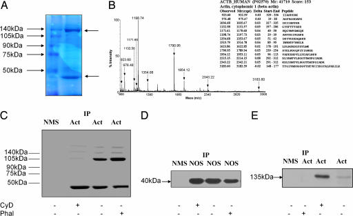
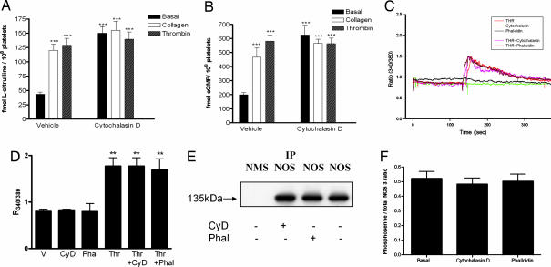
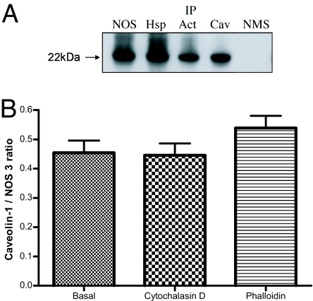
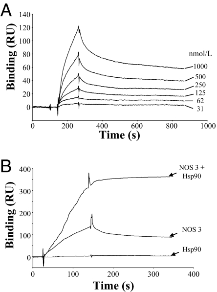
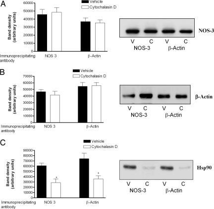
Cytoskeletal proteins are crucial in maintaining cellular structure and, in certain cell types, also play an essential role in motility and shape change. Nitric oxide (NO) is an important paracrine mediator of vascular and platelet function and is produced in the vasculature by the enzyme NO synthase type 3 (NOS-3). Here, we demonstrate in human platelets that the polymerization state of beta-actin crucially regulates the activation state of NOS-3, and hence NO formation, through altering its binding of heat shock protein 90 (Hsp90). We found that NOS-3 binds to the globular, but not the filamentous, form of beta-actin, and the affinity of NOS-3 for globular beta-actin is, in turn, increased by Hsp90. Formation of this ternary complex among NOS-3, globular beta-actin, and Hsp90, in turn, results in an increase in both NOS activity and cyclic guanosine-3',5'-monophosphate, an index of bioactive NO, as well as an increased rate of Hsp90 degradation, thus limiting the duration for which NOS-3 remains activated. These observations suggest that beta-actin plays a critical role in regulating NO formation and signaling in platelets.


Conflict of interest statement

The authors declare no conflict of interest.

Figures

Fig. 1.
Fig. 1.
NOS-3 binds to the globular, but not the filamentous, form of β-actin in platelets. (A) A protein of molecular mass 40 kDa coimmunoprecipitates with NOS-3. Human platelets, isolated from blood of healthy subjects (n = 4), were lysed, and NOS-3 was immunoprecipitated. This immunoprecipitate was subjected to SDS/PAGE on a 10% polyacrylamide gel, which was stained with Coomassie Brilliant blue. The left lane shows molecular mass markers, and the right lane is NOS-3 immunoprecipitate from platelets. Two bands predominate (arrowed), one at 135 kDa (the known molecular mass of NOS-3) and another at 40 kDa. (B) The 40-kDa protein is β-actin. The 40-kDa band was excised from the gel and digested with trypsin, and the digest analyzed by MALDI-TOF. The peptide mass fingerprint is that of β-actin. (C) β-Actin exists in both globular and filamentous forms in platelets. Human platelets, isolated from blood of healthy subjects (n = 6), were treated with vehicle, cytochalasin D 5 μmol/liter or phalloidin 5 μmol/liter, and lysed, and β-actin was immunoprecipitated from cell lysates. This immunoprecipitate was subjected to nondenaturing PAGE, followed by Western blotting for β-actin. Two predominant bands are seen, at 40 kDa (representing globular β-actin) and 105 kDa, as well as weaker bands at higher molecular mass. Treatment with cytochalasin D increases the density of the 40-kDa band and reduces the density of higher-molecular-mass bands. By contrast, treatment with phalloidin decreases the density of the 40-kDa band while increasing the density of higher-molecular-mass bands, especially the 105-kDa band. (D) NOS-3 binds globular, but not filamentous, β-actin. NOS-3 was immunoprecipitated from platelet lysates obtained from healthy subjects (n = 6), run on nondenaturing PAGE, and Western blotted for β-actin. Only a 40-kDa band was seen, with no evidence of higher-molecular-mass forms of β-actin present in the NOS-3 immunoprecipitates. Cytochalasin D increases, and phalloidin decreases, the density of the 40-kDa band. (E) Immunoprecipitation of β-actin pulls down NOS-3. β-Actin was immunoprecipitated from platelet lysates obtained from healthy subjects (n = 6) and was then run on SDS/PAGE and Western blotted for NOS-3. A 135-kDa band is seen, corresponding to the known molecular mass of NOS-3. Cytochalasin D increases, and phalloidin decreases, the density of the 135-kDa band. IP, immunoprecipitation; NMS, normal mouse serum; Act, anti-β-actin antibody; NOS, anti-NOS-3 antibody; CyD, cytochalasin D 5 μmol/liter; Phal, phalloidin 5 μmol/liter.
Fig. 2.
Fig. 2.
Polymerization status of β-actin regulates NOS-3 activity and bioactive NO, with no change in intracellular Ca2+ or serine phosphorylation of NOS-3. (A) NOS activity was determined, from l-[3H]arginine to l-[3H]citrulline conversion, in platelets isolated from healthy human subjects treated with cytochalasin D 5 μmol/liter or vehicle, which were coincubated with collagen 0.8 mg/liter, thrombin 1 unit/ml or corresponding vehicle. In platelets not treated with cytochalasin D, collagen and thrombin increase NOS activity. In platelets treated with cytochalasin D, basal NOS activity increases, and no further increase occurs with collagen or thrombin cotreatment. (n = 6, ± SD, ∗∗∗, P < 0.001 as compared with basal NOS activity in untreated platelets). (B) cGMP was assayed in platelets isolated from healthy human subjects treated with cytochalasin D 5 μmol/liter or vehicle, which were coincubated with collagen 0.8 mg/liter, thrombin 1 unit/ml or corresponding vehicle. In platelets not treated with cytochalasin D, collagen and thrombin increase intraplatelet cGMP. In platelets treated with cytochalasin D, basal cGMP is increased, and no further increase occurs with collagen or thrombin cotreatment. (n = 6, ± SD, ∗∗∗, P < 0.001 as compared with basal cGMP in untreated platelets). (C) A typical experiment showing changes in intraplatelet Ca2+ levels, as determined by fura-2 fluorescence and expressed as the ratio of emission at 510 nm after excitation at 340 and 380 nm (R340/380); tracings show no effect of cytochalasin D 5 μmol/liter or phalloidin 5 μmol/liter, whereas thrombin 1 unit/ml used as a positive control elicits an increase in intracellular Ca2+. The combination of thrombin with either cytochalasin D or phalloidin yields traces not different to that seen with thrombin alone. (D) Accumulated results showing effects of cytochalasin D (CyD), phalloidin (Phal) and thrombin (Thr), either alone or in combination as indicated, as compared with vehicle (V), on intraplatelet Ca2+, as determined from R340/380 (n = 6, ± SD, ∗∗, P < 0.01 as compared with vehicle). (E) Phosphoserine-modified NOS-3 was detected by Western blotting for phosphoserine in NOS-3 immunoprecipitates. IP, immunoprecipitation; NMS, normal mouse serum; NOS, anti-NOS-3 antibody; CyD, cytochalasin D 5 μmol/liter; Phal, phalloidin 5 μmol/liter. (F) Accumulated results showing phosphoserine-modified NOS-3, quantitated as the ratio of phosphoserine to NOS-3 bands as determined by scanning densitometry (n = 6, ±SD).
Fig. 3.
Fig. 3.
Hsp90 colocalizes with NOS-3 and β-actin. β-Actin, Hsp90, and NOS-3 were each detected (A–C, respectively) in fixed platelets from six healthy subjects, by ImmunoGold labeling with electron microscopy. β-Actin shows more extensive labeling than either Hsp90 or NOS-3, and this is distributed throughout the cytoplasm, whereas the labeling for Hsp90 and NOS-3 is clustered. Triple labeling shows multiple areas (arrowed) where β-actin (5-nm gold particles), NOS-3 (10-nm gold particles), and Hsp90 (15-nm gold particles) colocalize (D). g, platelet granule.
Fig. 4.
Fig. 4.
Hsp90, NOS-3 and β-actin localize to caveolae. (A) Platelets isolated from healthy subjects were lysed and, after immunoprecipitation of NOS-3 (NOS), Hsp90 (Hsp), β-actin (Act) or caveolin-1 (Cav) by using specific antibodies coated onto protein A-Sepharose beads, immunoprecipitates were run on SDS/PAGE by using a 12.5% polyacrylamide gel and Western blotted for caveolin-1. A 22-kDa protein is seen in all immunoprecipitates, which accords with the known molecular mass of caveolin-1. IP, immunoprecipitation; NMS, normal mouse serum used for immunoprecipitation as control. (B) Coimmunoprecipitation of caveolin-1 and NOS-3, quantitated as the ratio of caveolin-1 to NOS-3 bands as determined by scanning densitometry of Western blots of NOS-3 immunoprecpitates probed with anti-caveolin-1 or anti-NOS-3 antibody; cytochalasin D 5 μmol/liter and phalloidin 5 μmol/liter have no effect on caveolin-1 association to NOS-3 (n = 6, ±SD).
Fig. 5.
Fig. 5.
Globular β-actin forms a ternary complex with NOS-3 and Hsp90. (A) NOS-3 binds monomeric β-actin in a concentration-dependent manner. A CM5 sensor chip was chemically coated with purified monomeric β-actin [11,000 resonance units (RU)] and, after injection of different concentrations of recombinant NOS-3 (31 nmol/liter to 1 μmol/liter), binding was determined by surface plasmon resonance analysis. A representative experiment is shown from a total of three experiments. (B) Hsp90 increases binding of NOS-3 to monomeric β-actin. Binding was determined to monomeric β-actin immobilized on a CM5 sensor chip (13,000 RU), by surface plasmon resonance analysis, after preincubation of NOS-3 with Hsp90 in a 1:1 ratio. Although Hsp90 alone (500 nmol/liter) was found not to bind to monomeric β-actin, its preincubation with NOS-3 was found to increase the binding of NOS-3 by 2.8 ± 1.2-fold, and this binding became irreversible. A representative experiment is shown from a total of three experiments.
Fig. 6.
Fig. 6.
Formation of a ternary complex among Hsp90, β-actin, and NOS-3 leads to a decrease in bound Hsp90. Platelets from healthy subjects were treated with cytochalasin D 5 μmol/liter or vehicle, after which they were lysed and, after immunoprecipitation of NOS-3 or β-actin, immunoprecipitates were run on SDS/PAGE and Western blotted for either NOS-3 (A) or β-actin (B). Typical blots are shown (V, vehicle; C, cytochalasin D treatment), and results are presented for band density, as determined by scanning densitometry (n = 6, ±SD). Cytochalasin D does not affect the degree of coimmunoprecipitation of NOS-3 and β-actin. (C) Cytochalasin D treatment decreases Hsp90 levels in NOS-3 and β-actin immunoprecipitates. Platelets from healthy subjects were treated with cytochalasin D or vehicle, after which they were lysed and, after immunoprecipitation of NOS-3 or β-actin, immunoprecipitates were run on SDS/PAGE and Western blotted for Hsp90. A typical blot is shown (V, vehicle; C, cytochalasin D treatment), and results are presented for band density, as determined by scanning densitometry (n = 6, ±SD, ∗, P < 0.01 for cytochalasin D- versus vehicle-treated platelets).

Similar articles

Cited by

References

    1. Radomski MW, Moncada S. Adv Exp Med Biol. 1993;344:251–264. - PubMed
    1. Kinlay S, Ganz P. Am J Cardiol. 1997;80:11I–16I. - PubMed
    1. Ross R. N Engl J Med. 1999;340:115–126. - PubMed
    1. Anderson TJ. Heart Fail Rev. 2003;8:71–86. - PubMed
    1. Radomski MW, Palmer RM, Moncada S. Lancet. 1987;7:1057–1058. - PubMed

Publication types

LinkOut - more resources