

Photodynamic molecular beacon as an activatable photosensitizer based on protease-controlled singlet oxygen quenching and activation
- PMID: 17502620
- PMCID: PMC1868591
- DOI: 10.1073/pnas.0611142104
Photodynamic molecular beacon as an activatable photosensitizer based on protease-controlled singlet oxygen quenching and activation
Abstract
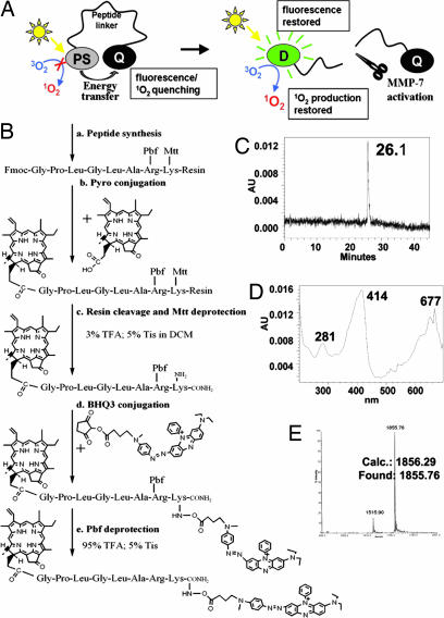
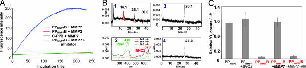
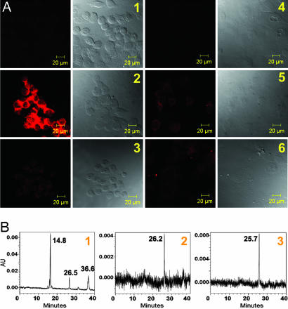
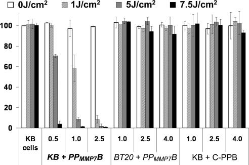
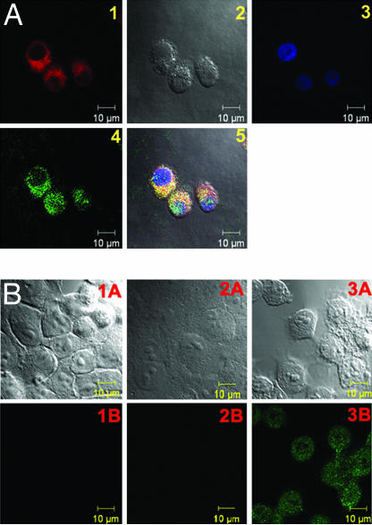
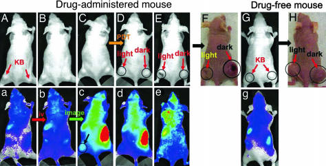
Molecular beacons are FRET-based target-activatable probes. They offer control of fluorescence emission in response to specific cancer targets, thus are useful tools for in vivo cancer imaging. Photodynamic therapy (PDT) is a cell-killing process by light activation of a photosensitizer (PS) in the presence of oxygen. The key cytotoxic agent is singlet oxygen ((1)O(2)). By combining these two principles (FRET and PDT), we have introduced a concept of photodynamic molecular beacons (PMB) for controlling the PS's ability to generate (1)O(2) and, ultimately, for controlling its PDT activity. The PMB comprises a disease-specific linker, a PS, and a (1)O(2) quencher, so that the PS's photoactivity is silenced until the linker interacts with a target molecule, such as a tumor-associated protease. Here, we report the full implementation of this concept by synthesizing a matrix metalloproteinase-7 (MMP7)-triggered PMB and achieving not only MMP7-triggered production of (1)O(2) in solution but also MMP7-mediated photodynamic cytotoxicity in cancer cells. Preliminary in vivo studies also reveal the MMP7-activated PDT efficacy of this PMB. This study validates the core principle of the PMB concept that selective PDT-induced cell death can be achieved by exerting precise control of the PS's ability to produce (1)O(2) by responding to specific cancer-associated biomarkers. Thus, PDT selectivity will no longer depend solely on how selectively the PS can be delivered to cancer cells. Rather, it will depend on how selective a biomarker is to cancer cells, and how selective the interaction of PMB is to this biomarker.
Conflict of interest statement
The authors declare no conflict of interest.
Figures
Similar articles
-
New photodynamic molecular beacons (PMB) as potential cancer-targeted agents in PDT.Bioorg Med Chem. 2018 Feb 1;26(3):688-702. doi: 10.1016/j.bmc.2017.12.034. Epub 2017 Dec 24. Bioorg Med Chem. 2018. PMID: 29338907
-
A tumor mRNA-triggered photodynamic molecular beacon based on oligonucleotide hairpin control of singlet oxygen production.Photochem Photobiol Sci. 2008 Jul;7(7):775-81. doi: 10.1039/b800653a. Epub 2008 Jun 9. Photochem Photobiol Sci. 2008. PMID: 18597024
-
Using the singlet oxygen scavenging property of carotenoid in photodynamic molecular beacons to minimize photodamage to non-targeted cells.Photochem Photobiol Sci. 2007 Dec;6(12):1311-7. doi: 10.1039/b706820d. Epub 2007 Aug 20. Photochem Photobiol Sci. 2007. PMID: 18046487
-
Advanced Photosensitizer Activation Strategies for Smarter Photodynamic Therapy Beacons.Angew Chem Int Ed Engl. 2019 Feb 25;58(9):2558-2569. doi: 10.1002/anie.201805246. Epub 2018 Dec 28. Angew Chem Int Ed Engl. 2019. PMID: 29890024 Review.
-
Modulation of photosensitization processes for an improved targeted photodynamic therapy.Curr Med Chem. 2010;17(32):3925-43. doi: 10.2174/092986710793205453. Curr Med Chem. 2010. PMID: 20858211 Review.
Cited by
-
Tumor-Specific Drug Release and Reactive Oxygen Species Generation for Cancer Chemo/Chemodynamic Combination Therapy.Adv Sci (Weinh). 2019 Jan 18;6(5):1801986. doi: 10.1002/advs.201801986. eCollection 2019 Mar 6. Adv Sci (Weinh). 2019. PMID: 30886808 Free PMC article.
-
Targeted photodynamic therapy in head and neck squamous cell carcinoma: heading into the future.Lasers Med Sci. 2015 Dec;30(9):2381-7. doi: 10.1007/s10103-014-1703-4. Epub 2015 Jan 7. Lasers Med Sci. 2015. PMID: 25563461 Review.
-
Mesoporous semiconductors combined with up-conversion nanoparticles for enhanced photodynamic therapy under near infrared light.RSC Adv. 2019 Jun 3;9(30):17273-17280. doi: 10.1039/c9ra03116b. eCollection 2019 May 29. RSC Adv. 2019. PMID: 35519878 Free PMC article.
-
Smart Polymeric Delivery System for Antitumor and Antimicrobial Photodynamic Therapy.Front Bioeng Biotechnol. 2021 Nov 4;9:783354. doi: 10.3389/fbioe.2021.783354. eCollection 2021. Front Bioeng Biotechnol. 2021. PMID: 34805129 Free PMC article. Review.
-
Controllable Photodynamic Therapy Implemented by Regulating Singlet Oxygen Efficiency.Adv Sci (Weinh). 2017 Jun 23;4(7):1700113. doi: 10.1002/advs.201700113. eCollection 2017 Jul. Adv Sci (Weinh). 2017. PMID: 28725533 Free PMC article. Review.
References
-
- Pinthus JH, Bogaards A, Weersink R, Wilson BC, Trachtenberg J. J Urol. 2006;175:1201–1207. - PubMed
-
- Chen B, Pogue BW, Hasan T. Exp Opin Drug Del. 2005;2:477–487. - PubMed
-
- Sharman WM, van Lier JE, Allen CM. Adv Drug Del Rev. 2004;56:53–76. - PubMed
-
- Zhang M, Zhang Z, Blessington D, Li H, Busch TM, Madrak V, Miles J, Chance B, Glickson JD, Zheng G. Bioconjug Chem. 2003;14:709–714. - PubMed
Publication types
MeSH terms
Substances
Grants and funding
LinkOut - more resources
Full Text Sources
Other Literature Sources
