Comparative Study
. 2007 Jun 11;204(6):1303-10.
doi: 10.1084/jem.20062129. Epub 2007 May 14.

Cyclic adenosine monophosphate is a key component of regulatory T cell-mediated suppression

Affiliations
Comparative Study

Cyclic adenosine monophosphate is a key component of regulatory T cell-mediated suppression

Tobias Bopp et al. J Exp Med. .

Abstract

Naturally occurring regulatory T cells (T reg cells) are a thymus-derived subset of T cells, which are crucial for the maintenance of peripheral tolerance by controlling potentially autoreactive T cells. However, the underlying molecular mechanisms of this strictly cell contact-dependent process are still elusive. Here we show that naturally occurring T reg cells harbor high levels of cyclic adenosine monophosphate (cAMP). This second messenger is known to be a potent inhibitor of proliferation and interleukin 2 synthesis in T cells. Upon coactivation with naturally occurring T reg cells the cAMP content of responder T cells is also strongly increased. Furthermore, we demonstrate that naturally occurring T reg cells and conventional T cells communicate via cell contact-dependent gap junction formation. The suppressive activity of naturally occurring T reg cells is abolished by a cAMP antagonist as well as by a gap junction inhibitor, which blocks the cell contact-dependent transfer of cAMP to responder T cells. Accordingly, our results suggest that cAMP is crucial for naturally occurring T reg cell-mediated suppression and traverses membranes via gap junctions. Hence, naturally occurring T reg cells unexpectedly may control the immune regulatory network by a well-known mechanism based on the intercellular transport of cAMP via gap junctions.


Figures
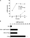
Figure 1.
Figure 1.
Naturally occurring T reg cell–mediated suppression is accompanied by a strong increase of cAMP in the suppressed CD4+ T cells. (A) CFSE-labeled CD4+ T cells (CD4; ♦) and naturally occurring T reg cells (▪) were stimulated alone or in co-culture with anti-CD3 mAb and A20 cells as accessory cells for 4, 8, and 16 h. At the indicated time points, the CD4+ T cells from the co-culture were reisolated using FACS-based cell sorting (CD4 (+nTreg); ▴) to a purity >99%. The cytosolic cAMP concentration was assessed using a cAMP-specific ELISA. One representative result of two independent experiments is shown. (B) CD4+ T cells and naturally occurring T reg cells were activated as described in A. Alternatively, CD4+ T cells were stimulated in the presence of 1 μM forskolin. After 20 h of stimulation, cytosolic cAMP concentrations were measured. Shown are the means ± SD of three independent experiments each performed in triplicates.
Figure 2.
Figure 2.
cAMP is a key component of naturally occurring T reg cell–mediated suppression. CD4+ T cells were preincubated for 30 min either with the specific cAMP antagonist Rp-cAMPS or its solvent as described in Materials and methods. Upon preincubation, cells were washed and activated separately (CD4) or in co-culture with naturally occurring T reg cells (CD4 +nTreg) at the indicated ratios. After 20 h of stimulation quantitative RT-PCR for the expression of IL-2 mRNA was performed and data were normalized according to the expression of EF1α mRNA and expressed as a percentage of IL-2 mRNA by setting the relative IL-2 mRNA expression of the CD4 single culture to 100%. One representative assay performed in triplicates ± SD is shown.
Figure 3.
Figure 3.
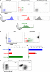
Naturally occurring T reg cells and conventional CD4+ T cells communicate via GJ in vivo and in vitro. (A) OT-II, CD90.2-positive, RAG-2–deficient mice were immunized subcutaneously with OVA323-339 in the left hind footpad. 6 h later vehicle or calcein-loaded naturally occurring T reg cells isolated from the spleens of CD90.1-positive congenic OT-II mice and preactivated for 48 h in vitro were injected i.v. 24 h after immunization, the draining and the nondraining LNs were dissected and LN cells were stained for the expression of CD4, CD69, and CD90.1. The intracellular calcein-level (A) and the expression of CD69 (B) were analyzed by gating on propidium iodide–negative and CD4+ LN cells. (C) CD4+ T cells from BALB/c mice (H2-Kd) were stimulated in vitro in the absence (single-culture; blue) or presence of calcein-loaded naturally occurring T reg cells isolated from the spleens of C57BL/6 (H2-Kb). After 20 h, cells were stained with an anti–H2-Kb mAb and GJIC was assessed by analyzing calcein transfer (red, calcein-low; green, calcein-high). (D and E) Simultaneously, CD4+ T cells from the single culture and H2-Kb-negative, calcein-high (green) or calcein-low (red) CD4+ T cells were reisolated from the co-cultures using FACS-based cell sorting and analyzed for their cytosolic cAMP concentration and the expression of IL-2 mRNA. (C–E) One representative result of three independent experiments. (F) Conventional CD90.1-positive CD4+ T cells were stimulated in the presence of CD90.2-positive naturally occurring T reg cells or CD90.2-positive naturally occurring T reg cells that had been transfected with Alexa Fluor 488 cAMP. After 20 h, cells were stained for expression of CD90.1 to discriminate CD4+ T cells and T reg cells and analyzed for the diffusion of Alexa Fluor 488 cAMP to CD90.1-positive CD4+ T cells.
Figure 4.
Figure 4.
Blocking GJIC simultaneously reduces the influx of calcein and the inhibition of IL-2 expression in CD4+ T cells upon contact with naturally occurring T reg cells. (A) CD4+ T cells from BALB/c mice (H2-Kd) were activated separately (CD4) or in co-culture with calcein-loaded naturally occurring T reg cells (CD4 +nTreg) from C57BL/6 mice (H2-Kb). As depicted, the cells were stimulated in the presence or absence of the GJ mimetic peptide GAP27 or a scrambled control peptide SC-GAP27, consisting of the same amino acids in a different order. 20 h upon stimulation, the calcein transfer was assessed by gating on CD4+ T cells (H2-Kb negative, propidium iodide negative). (B) In parallel, the CD4+ T cells were reisolated from the co-culture using FACS-based cell sorting to a purity >99%. Expression of IL-2 mRNA was assessed in triplicates by quantitative RT-PCR and data were normalized according to the expression of EF1α mRNA. Shown are the means ± SD of three independent experiments. N.d., not detectable.

Similar articles

Cited by

References

    1. Gershon, R.K., and K. Kondo. 1970. Cell interactions in the induction of tolerance: the role of thymic lymphocytes. Immunology. 18:723–737. - PMC - PubMed
    1. Sakaguchi, S., N. Sakaguchi, M. Asano, M. Itoh, and M. Toda. 1995. Immunologic self-tolerance maintained by activated T cells expressing IL-2 receptor alpha-chains (CD25). Breakdown of a single mechanism of self-tolerance causes various autoimmune diseases. J. Immunol. 155:1151–1164. - PubMed
    1. Sakaguchi, S. 2004. Naturally arising CD4+ regulatory t cells for immunologic self-tolerance and negative control of immune responses. Annu. Rev. Immunol. 22:531–562. - PubMed
    1. Piccirillo, C.A., J.J. Letterio, A.M. Thornton, R.S. McHugh, M. Mamura, H. Mizuhara, and E.M. Shevach. 2002. CD4+CD25+ regulatory T cells can mediate suppressor function in the absence of transforming growth factor β1 production and responsiveness. J. Exp. Med. 196:237–246. - PMC - PubMed
    1. Fahlen, L., S. Read, L. Gorelik, S.D. Hurst, R.L. Coffman, R.A. Flavell, and F. Powrie. 2005. T cells that cannot respond to TGF-β escape control by CD4+CD25+ regulatory T cells. J. Exp. Med. 201:737–746. - PMC - PubMed

Publication types