Comparative Study
. 2007 Jun 11;204(6):1267-72.
doi: 10.1084/jem.20062497. Epub 2007 May 14.

RANK signals from CD4(+)3(-) inducer cells regulate development of Aire-expressing epithelial cells in the thymic medulla

Affiliations
Comparative Study

RANK signals from CD4(+)3(-) inducer cells regulate development of Aire-expressing epithelial cells in the thymic medulla

Simona W Rossi et al. J Exp Med. .

Abstract

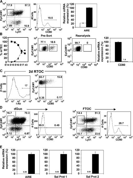
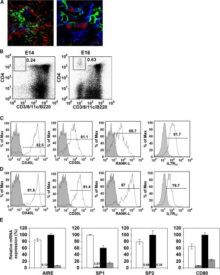
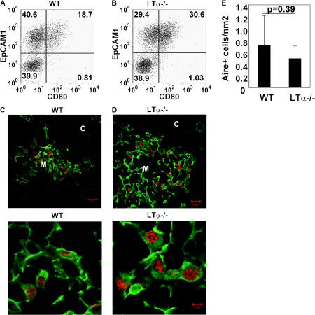
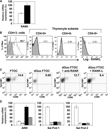
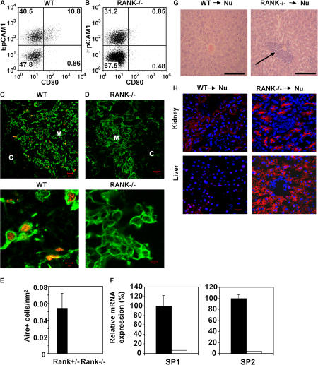
Aire-expressing medullary thymic epithelial cells (mTECs) play a key role in preventing autoimmunity by expressing tissue-restricted antigens to help purge the emerging T cell receptor repertoire of self-reactive specificities. Here we demonstrate a novel role for a CD4(+)3(-) inducer cell population, previously linked to development of organized secondary lymphoid structures and maintenance of T cell memory in the functional regulation of Aire-mediated promiscuous gene expression in the thymus. CD4(+)3(-) cells are closely associated with mTECs in adult thymus, and in fetal thymus their appearance is temporally linked with the appearance of Aire(+) mTECs. We show that RANKL signals from this cell promote the maturation of RANK-expressing CD80(-)Aire(-) mTEC progenitors into CD80(+)Aire(+) mTECs, and that transplantation of RANK-deficient thymic stroma into immunodeficient hosts induces autoimmunity. Collectively, our data reveal cellular and molecular mechanisms leading to the generation of Aire(+) mTECs and highlight a previously unrecognized role for CD4(+)3(-)RANKL(+) inducer cells in intrathymic self-tolerance.


Figures

Figure 1.
Figure 1.
Haemopoietic cells regulate mTEC development. EpCAM1+Ly51 mTECs in 7 d FTOCs (A) can be subdivided into CD80 and CD80+ subsets. qPCR anaysis shows mRNA for Aire is abundant in CD80+ mTECs (black bar) compared with CD80 mTECs (white bar). The left graph in B shows percentages of CD80 mTECs (▪) and CD80+ (▴) mTEC subsets within the total mTEC population, calculated after flow cytometric analysis of digested thymuses of the indicated ages. H-2b CD80 mTECs, shown by FACS (B) to lack surface CD80 expression and by PCR to lack CD80 mRNA (black bars, CD80+ mTEC; white bars, CD80 mTECs), were used to make RTOCs with H-2d thymus suspensions. RTOCs were analyzed for I-Ab (C, left), Ly51, EpCAM1, and CD80 expression after 2 d. Gating on I-Ab+ mTECs (C, right) shows CD80 mTECs have generated CD80+ mTECs. Analysis of mTECs in FTOCs or dGuo-treated FTOCs (D) shows absence of the CD80+ mTEC subset in dGuo FTOCs. qPCR analysis (E) shows Aire, SP1, and SP2 expression in mTECs from FTOCs (black bars) but not dGuo-treated FTOCs (white bars).
Figure 2.
Figure 2.
CD4+3 LTi cells regulate mTEC development. Thymic sections of 4-wk-old Rag1−/− mice (A, left) were stained with antibodies to mTECs (ERTR5, red), CD4 (green), CD3 (white), and CD11c (blue). In the right panel of A, sections were stained for Aire (red), CD4 (green), and keratin5+ mTECs (blue). In both cases, CD4+3 cells appear green. Bars, 5 μm. Flow cytometry of cells harvested from FTOCs from E14 and E16 WT embryos (B) shows CD4+3 cells that lack expression of CD8, CD11c, and B220, whereas CD4+3 cells from adult thymus (C) express OX40L, CD30L, IL-7Rα, and RANKL in a manner comparable to splenic CD4+3 cells (D). Shaded histograms are staining controls. E shows qPCR analysis of mTECs in RTOCs initiated from either dGuo-treated stroma alone (vertical bars), or with added CD4+3 LTi cells (E, black bars) or CD4+8+ thymocytes (E, hatched bars). Expression levels in unmanipulated FTOCs are shown for comparison (E, white bars.)
Figure 3.
Figure 3.
Aire+ mTECs are present in LTα−/− mice. Stroma-enriched adult thymus digests from WT (A) and LTα−/− mice (B) were stained with antibodies to CD45, EpCAM1, Ly51, and CD80. Data are gated on CD45Ly51 cells. Sections of WT (C) and LTα−/− (D) adult thymus were stained for keratin 5 (green) and Aire (red). The bottom two panels show higher power magnification. Bars: (C and D, top) 20 μm; (C and D, bottom) 5 μm. E shows quantitation of Aire+ mTECs in sections of WT and LTα−/− mice. Student's t test was run to determine the p-value, which was not significant.
Figure 4.
Figure 4.
RANK–RANKL interactions promote Aire+ mTEC development. qPCR analysis of RANK expression (A) in EpCAM1+Ly51 mTECs (black bar) and EpCAM1+Ly51+ cTEC (white bar). B shows analysis of RANKL expression in thymic CD4+3 LTi cells and thymocyte subsets. Shaded histograms are staining controls. C shows analysis of EpCAM1+Ly51CD80+ mTEC in FTOCs and dGuo-treated FTOCs cultured in the absence or presence of anti-RANK or recombinant RANKL for 2 d. Quadrant gates are set using staining levels obtained using isotype-matched control antibodies. (D) qPCR analysis of Aire, SP1, and SP2 expression was performed in untreated FTOCs (white bars) dGuo FTOC (vertical bars), and in dGuo FTOCs treated with either anti-RANK (black bars) or recombinant RANKL (hatched bars).
Figure 5.
Figure 5.
Lack of Aire+CD80+ mTECs in RANK−/− mice promotes autoimmunity. Stroma-enriched adult thymus digests from WT (A) and RANK−/− mice (B) were stained for CD45, EpCAM1, Ly51, and CD80. Data are gated on CD45Ly51 cells. In C and D, tissue sections of WT and RANK−/− adult thymus were stained for keratin 5 (green) and Aire (red). Higher power magnification is shown in the bottom panels. Bars: (C and D, top) 20 μm; (C and D, bottom) 5 μm. E shows quantitation of Aire+ mTECs in sections of RANK+/− and RANK−/− thymus. F is qPCR highlighting absence of TRA expression in RANK−/− thymus (white bars) compared with WT thymus (black bars). Histological analysis of leukocytic infiltrates in the liver of nude mice (arrow) transplanted with RANK−/− dGuo-treated, but not WT thymus, is in G. (H) Immunohistochemical analysis of Rag−/− tissues using serum obtained from RANK−/− and WT transplanted nude mice. Red, autoantibody staining; blue, DAPI staining. Bars: G, 100 μm; H, 20 μm.

References

    1. Kyewski, B., and L. Klein. 2006. A central role for central tolerance. Annu. Rev. Immunol. 24:571–606. - PubMed
    1. Ueno, T., F. Saito, D.H. Gray, S. Kuse, K. Hieshima, H. Nakano, T. Kakiuchi, M. Lipp, R.L. Boyd, and Y. Takahama. 2004. CCR7 signals are essential for cortex-to-medulla migration of developing thymocytes. J. Exp. Med. 200:493–505. - PMC - PubMed
    1. Derbinski, J., A. Schulte, B. Kyewski, and L. Klein. 2001. Promiscuous gene expression in medullary thymic epithelial cells mirrors the peripheral self. Nat. Immunol. 2:1032–1039. - PubMed
    1. Anderson, M.S., E.S. Venanzi, L. Klein, Z. Chen, S.P. Berzins, S.J. Turley, H. von Boehmer, R. Bronson, A. Dierich, C. Benoist, and D. Mathis. 2002. Projections of an immunological self shadow within the thymus by the aire protein. Science. 298:1395–1401. - PubMed
    1. Devoss, J., Y. Hou, K. Jahannes, W. Lu, G.I. Liou, J. Rinn, H. Chang, R. Caspi, L. Fong, and M.S. Anderson. 2006. Spontaneous autoimmunity prevented by thymic expression of a single self-antigen. J. Exp. Med. 203:2727–2735. - PMC - PubMed

Publication types

MeSH terms