. 2007 Jun 11;204(6):1257-65.
doi: 10.1084/jem.20062512. Epub 2007 May 14.

Adenosine generation catalyzed by CD39 and CD73 expressed on regulatory T cells mediates immune suppression

Affiliations

Adenosine generation catalyzed by CD39 and CD73 expressed on regulatory T cells mediates immune suppression

Silvia Deaglio et al. J Exp Med. .

Abstract

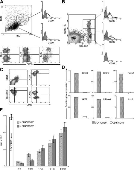
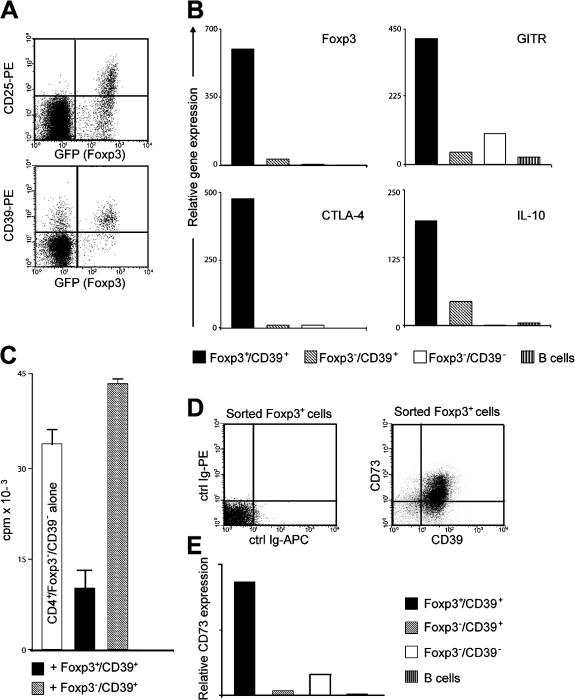
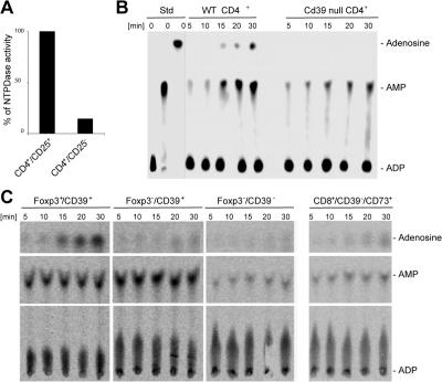
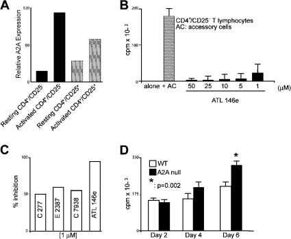
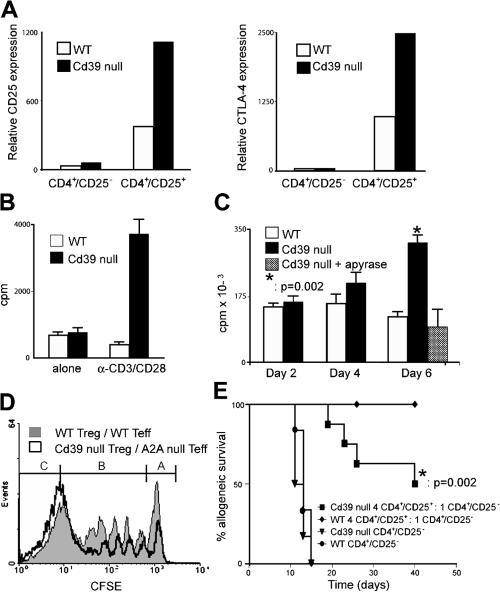
The study of T regulatory cells (T reg cells) has been limited by the lack of specific surface markers and an inability to define mechanisms of suppression. We show that the expression of CD39/ENTPD1 in concert with CD73/ecto-5'-nucleotidase distinguishes CD4(+)/CD25(+)/Foxp3(+) T reg cells from other T cells. These ectoenzymes generate pericellular adenosine from extracellular nucleotides. The coordinated expression of CD39/CD73 on T reg cells and the adenosine A2A receptor on activated T effector cells generates immunosuppressive loops, indicating roles in the inhibitory function of T reg cells. Consequently, T reg cells from Cd39-null mice show impaired suppressive properties in vitro and fail to block allograft rejection in vivo. We conclude that CD39 and CD73 are surface markers of T reg cells that impart a specific biochemical signature characterized by adenosine generation that has functional relevance for cellular immunoregulation.


Figures

Figure 1.
Figure 1.
CD4+/CD25+ cells express CD39. (A) The expression of CD39 (open profiles) on mouse lymph node cell suspensions is shown in the histograms on the right, contrasted against control IgG (gray). The expression of CD39 on CD4+, CD8+, and B220+ subsets is shown in the density plots. (B) Lymph node cells were stained with CD4-Cy5 and CD25-PE. CD39 expression in gated populations is shown in the histograms (open) with irrelevant control (gray). (C) CD4+ cells were stained for CD39 (horizontal axis) and the indicated markers. (D) Real-time PCR analysis in CD4+/CD39+ and CD4+/CD39 cells. (E) 5 × 10⁴ CD4+/CD25 T cells were mixed with CD4+/CD39+ or CD4+/CD25+ cells. [3H]Thymidine incorporation was measured after 3 d. Data are means with SEM (error bars) of three independent experiments.
Figure 2.
Figure 2.
Foxp3+ cells express CD39. (A) CD4+ T cells from Foxp3+ (GFP+) knock-in mice stained with CD25-PE or CD39-PE. (B) Real-time PCR analysis of Foxp3+/CD39+ (black bars), Foxp3/CD39+ (shaded bars), and Foxp3/CD39 (white bars) cells. (C) Foxp3+/CD39+ suppresses T effector (Foxp3/CD39 CD4+) proliferation. [3H]Thymidine incorporation was measured after 72 h. Data are representative of two independent experiments, with error bars representing the SD of triplicate wells. (D) Foxp3+(GFP+) cells stained for CD39-APC and CD73-PE. (E) CD73 mRNA expression in Foxp3+/CD39+ (black bars), Foxp3/CD39+ (shaded bars), Foxp3/CD39 (white bars), and B lymphocytes (striped bars).
Figure 3.
Figure 3.
Concomitant expression of CD39/CD73 and generation of adenosine distinguishes T reg cells from other T cells. (A) CD39/NTPDase activity in CD4+/CD25+ and CD4+/CD25 cells. (B) Hydrolysis of extracellular 14C-radiolabeled ADP to adenosine is catalyzed by WT CD4+ (lanes 4–8) but not Cd39-null CD4+ (lanes 9–13) mouse T cells. The radiolabeled standards are designated as ADP, AMP, and Adenosine (lanes 1–3). (C) Hydrolysis of extracellular ADP by Foxp3+/CD39+ (first to fifth lanes), Foxp3/CD39+ (6th to 10th lanes), and Foxp3/CD39 (11th to 15th lanes) cells.
Figure 4.
Figure 4.
Adenosine generated by CD39 and CD73 suppresses T cell proliferation. (A) mRNA expression of adenosine A2A receptor. Cells were activated for 5 d in vitro. Data are means of three independent experiments. (B) ATL146e inhibits T cell proliferation in response to alloantigens. 5 × 10⁴ CD4+/CD25 T cells were cultured alone or in the presence of irradiated allogeneic splenocytes (shaded bar) with ATL146e at indicated concentrations. Data are means with SEM (error bars) of more than five independent experiments (P < 0.05 vs. CD4+/CD25 cells alone). (C) Inhibition of T cell proliferation by adenosine receptor agonists. C277, E2387, C7938, and ATL146e (1 μM final) were added at the beginning of the cultures. [3H]Thymidine incorporation was assayed at day 5. Data are representative of five independent experiments. (D) Proliferation of CD4+/CD25 T cells purified from A2A-null (black bars) or WT (white bars) mice and stimulated by 2.5 μg/ml of plate-bound anti-CD3 and 2.5 μg/ml of soluble anti-CD28. [3H]Thymidine was added for the last 8 h of culture. Representative data are from three experiments. *, P < 0.002 between WT and A2A null at 6 d of culture.
Figure 5.
Figure 5.
T reg cells from Cd39-null mice are constitutively activated and fail to suppress CD4+/CD25+ cell proliferation. (A) CD25 and CTLA-4 mRNA expression in CD4+/CD25 and CD4+/CD25+ cells isolated from Cd39-null (black bars) or WT mice (white bars). (B) Proliferation assay of CD4+/CD25+ T cells purified from Cd39-null (black bars) or WT (white bars) mice. Cells were cultured in the presence or absence of 2.5 μg/ml of plate-bound anti-CD3 and 2.5 μg/ml of soluble anti-CD28 for 3 d. (C) Proliferation of CD4+/CD25 T cells purified from Cd39-null (black bars) or WT (white bars) mice cultured in the presence of irradiated allogeneic splenocytes. Apyrase reconstitution effects are noted at day 6 (shaded bars). Error bars represent the SEM of three independent experiments. (D) T reg cell function. Cd39-null T reg cell effects of the stimulation of A2A-null Teff (open) and WT T reg cells on WT Teff (closed; 1:1 ratio) are compared at day 5. A represents the nonproliferating cell populations, B represents CD4+/CD25 cells that have entered into the cell cycle, and C represents unlabeled T reg cells. The percentage of cells present in A or B was quantified using FlowJo software. Data are representative of three independent experiments. (E) Skin allograft survival. C57BL/6 Rag 1–deficient mice received allogeneic skin grafts 24 h after the passive transfer of CD4+/CD25+ and/or CD4+/CD25 cells from WT or Cd39-null mice in a ratio of 4:1 (32, 33). Mice receiving WT (circles) or Cd39-null (triangles) CD4+/CD25 cells rejected the skin graft at a median of 13 d (n = 6). Mice receiving WT CD4+/CD25+ cells transferred in excess (diamonds; 4:1) with WT CD4+/CD25 cells showed long-term graft survival (n = 9). Four out of eight mice receiving Cd39-null CD4+/CD25+ cells transferred in excess (squares; 4:1) with Cd39-null CD4+/CD25 cells rejected the skin allograft at a median of 24.5 d (P = 0.02).

Similar articles

Cited by

  • NF-κB: roles and regulation in different CD4(+) T-cell subsets.
    Oh H, Ghosh S. Oh H, et al. Immunol Rev. 2013 Mar;252(1):41-51. doi: 10.1111/imr.12033. Immunol Rev. 2013. PMID: 23405894 Free PMC article. Review.
  • The CD39-adenosinergic axis in the pathogenesis of renal ischemia-reperfusion injury.
    Roberts V, Lu B, Rajakumar S, Cowan PJ, Dwyer KM. Roberts V, et al. Purinergic Signal. 2013 Jun;9(2):135-43. doi: 10.1007/s11302-012-9342-3. Epub 2012 Nov 29. Purinergic Signal. 2013. PMID: 23188420 Free PMC article. Review.
  • Classification of current anticancer immunotherapies.
    Galluzzi L, Vacchelli E, Bravo-San Pedro JM, Buqué A, Senovilla L, Baracco EE, Bloy N, Castoldi F, Abastado JP, Agostinis P, Apte RN, Aranda F, Ayyoub M, Beckhove P, Blay JY, Bracci L, Caignard A, Castelli C, Cavallo F, Celis E, Cerundolo V, Clayton A, Colombo MP, Coussens L, Dhodapkar MV, Eggermont AM, Fearon DT, Fridman WH, Fučíková J, Gabrilovich DI, Galon J, Garg A, Ghiringhelli F, Giaccone G, Gilboa E, Gnjatic S, Hoos A, Hosmalin A, Jäger D, Kalinski P, Kärre K, Kepp O, Kiessling R, Kirkwood JM, Klein E, Knuth A, Lewis CE, Liblau R, Lotze MT, Lugli E, Mach JP, Mattei F, Mavilio D, Melero I, Melief CJ, Mittendorf EA, Moretta L, Odunsi A, Okada H, Palucka AK, Peter ME, Pienta KJ, Porgador A, Prendergast GC, Rabinovich GA, Restifo NP, Rizvi N, Sautès-Fridman C, Schreiber H, Seliger B, Shiku H, Silva-Santos B, Smyth MJ, Speiser DE, Spisek R, Srivastava PK, Talmadge JE, Tartour E, Van Der Burg SH, Van Den Eynde BJ, Vile R, Wagner H, Weber JS, Whiteside TL, Wolchok JD, Zitvogel L, Zou W, Kroemer G. Galluzzi L, et al. Oncotarget. 2014 Dec 30;5(24):12472-508. doi: 10.18632/oncotarget.2998. Oncotarget. 2014. PMID: 25537519 Free PMC article. Review.
  • Age-Related Changes in Thymic Central Tolerance.
    Srinivasan J, Lancaster JN, Singarapu N, Hale LP, Ehrlich LIR, Richie ER. Srinivasan J, et al. Front Immunol. 2021 Apr 22;12:676236. doi: 10.3389/fimmu.2021.676236. eCollection 2021. Front Immunol. 2021. PMID: 33968086 Free PMC article. Review.
  • Regulatory Role of Immune Cell-Derived Extracellular Vesicles in Cancer: The Message Is in the Envelope.
    Li C, Donninger H, Eaton J, Yaddanapudi K. Li C, et al. Front Immunol. 2020 Jul 16;11:1525. doi: 10.3389/fimmu.2020.01525. eCollection 2020. Front Immunol. 2020. PMID: 32765528 Free PMC article. Review.

References

    1. Sakaguchi, S., N. Sakaguchi, M. Asano, M. Itoh, and M. Toda. 1995. Immunologic self-tolerance maintained by activated T cells expressing IL-2 receptor alpha-chains (CD25). Breakdown of a single mechanism of self-tolerance causes various autoimmune diseases. J. Immunol. 155:1151–1164. - PubMed
    1. Zheng, X.X., A. Sanchez-Fueyo, M. Sho, C. Domenig, M.H. Sayegh, and T.B. Strom. 2003. Favorably tipping the balance between cytopathic and regulatory T cells to create transplantation tolerance. Immunity. 19:503–514. - PubMed
    1. Sakaguchi, S. 2000. Regulatory T cells: key controllers of immunologic self-tolerance. Cell. 101:455–458. - PubMed
    1. Belkaid, Y., and B.T. Rouse. 2005. Natural regulatory T cells in infectious disease. Nat. Immunol. 6:353–360. - PubMed
    1. Rouse, B.T., and S. Suvas. 2004. Regulatory cells and infectious agents: detentes cordiale and contraire. J. Immunol. 173:2211–2215. - PubMed

Publication types