Review
. 2007:2007:30873.
doi: 10.1155/2007/30873. Epub 2007 Jan 16.

The role of the medial prefrontal cortex-amygdala circuit in stress effects on the extinction of fear

Affiliations
Review

The role of the medial prefrontal cortex-amygdala circuit in stress effects on the extinction of fear

Irit Akirav et al. Neural Plast. 2007.

Abstract

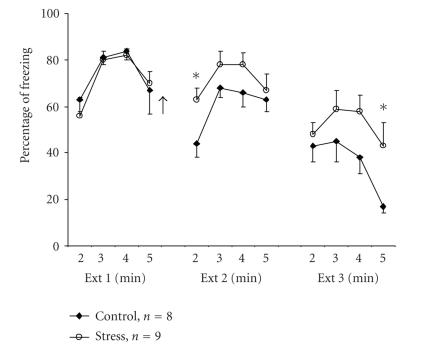
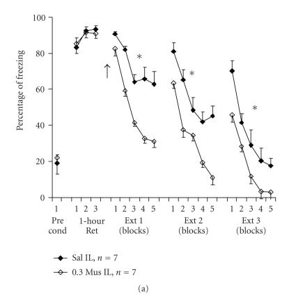
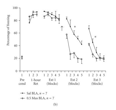
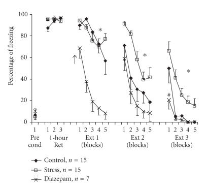
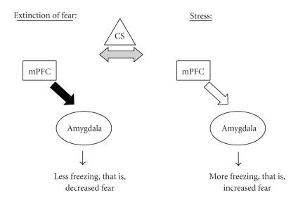
Stress exposure, depending on its intensity and duration, affects cognition and learning in an adaptive or maladaptive manner. Studies addressing the effects of stress on cognitive processes have mainly focused on conditioned fear, since it is suggested that fear-motivated learning lies at the root of affective and anxiety disorders. Inhibition of fear-motivated response can be accomplished by experimental extinction of the fearful response to the fear-inducing stimulus. Converging evidence indicates that extinction of fear memory requires plasticity in both the medial prefrontal cortex and the amygdala. These brain areas are also deeply involved in mediating the effects of exposure to stress on memory. Moreover, extensive evidence indicates that gamma-aminobutyric acid (GABA) transmission plays a primary role in the modulation of behavioral sequelae resulting from a stressful experience, and may also partially mediate inhibitory learning during extinction. In this review, we present evidence that exposure to a stressful experience may impair fear extinction and the possible involvement of the GABA system. Impairment of fear extinction learning is particularly important as it may predispose some individuals to the development of posttraumatic stress disorder. We further discuss a possible dysfunction in the medial prefrontal cortex-amygdala circuit following a stressful experience that may explain the impaired extinction caused by exposure to a stressor.


Figures

Figure 1
Figure 1
Stress impairs extinction of contextual fear conditioning. Rats were given 3 mild foot shocks in the conditioning chamber. On the next day, the rats were placed in the extinction (Ext) chamber for 5 minutes and no shock was administered (Ext 1; the last 4 minutes are presented since all animals showed high levels of freezing in the first minute). Immediately afterwards, the animals were returned to their home cage (control) or placed on an elevated platform for 30 minutes (stress). Animals were exposed to additional 5 minutes in the extinction chamber, without shocks, on days 3 (Ext 2) and 4 (Ext 3). The stressed animals showed significantly higher levels of freezing compared with the control group during the second minute of Ext 2 (∗; P < .05) and the fifth minute of Ext 3 (∗; P < .05). Arrow denotes time of exposure to stress.
Figure 2
Figure 2
(a) A low volume of muscimol microinfused into the infralimbic cortex before extinction training facilitates extinction learning. Rats received 7 pairings of a tone with a foot shock in the conditioning chamber. After 1 hour, three tones were delivered in the absence of foot shock (1-hour Ret). On the next day, the animals were microinfused with a total of 0.3 μl saline (Sal) or muscimol (0.3 Mus) to the infralimbic cortex (IL) and were exposed to 15 tones without foot shocks (Ext 1; presented as 5 blocks of 3 trials). Animals were exposed to additional 15 tones on days 4 (Ext 2) and 5 (Ext 3), without further administration of the drug. Muscimol IL animals showed significantly lower levels of freezing compared with the saline group in Ext 1 (∗; P < .001), Ext 2 (∗; P < .01) and Ext 3 (∗; P < .05). This supports a selective involvement of the IL in facilitating extinction of conditioned fear (see Akirav et al. [51]). Arrow denotes time of drug infusion. The Pre cond data points indicate the amount of freezing exhibited by rats prior to commencement of fear conditioning. (b) A low volume of muscimol microinfused to the basolateral amygdala following a short extinction training session facilitates extinction consolidation. Rats received 7 pairings of a tone with a foot shock in the conditioning chamber. After 1 hour, three tones were delivered in the absence of foot shock (1-hour Ret). On the next day, the animals underwent a short extinction training session consisting of 5 tones (Ext 1; presented as 5 trials), and were thereafter microinfused with a total volume of 0.5 μl saline (Sal) or muscimol (0.5 Mus) to the basolateral amygdala (BLA). On days 4 and 5 (Ext 2 and Ext 3, resp.), the animals were exposed to 15 tones without foot shocks (presented as 5 blocks of 3 trials). The BLA muscimol group showed significantly reduced levels of freezing compared with the other two groups during Ext 2 (∗; P < .001) and Ext 3 (∗; P < .05). This supports the selective involvement of the BLA in facilitating consolidation of extinction of conditioned fear (see Akirav et al. [51]). Arrow denotes time of drug infusion. The Pre cond data points indicate the amount of freezing exhibited by rats prior to commencement of fear conditioning.
Figure 2
Figure 2
(a) A low volume of muscimol microinfused into the infralimbic cortex before extinction training facilitates extinction learning. Rats received 7 pairings of a tone with a foot shock in the conditioning chamber. After 1 hour, three tones were delivered in the absence of foot shock (1-hour Ret). On the next day, the animals were microinfused with a total of 0.3 μl saline (Sal) or muscimol (0.3 Mus) to the infralimbic cortex (IL) and were exposed to 15 tones without foot shocks (Ext 1; presented as 5 blocks of 3 trials). Animals were exposed to additional 15 tones on days 4 (Ext 2) and 5 (Ext 3), without further administration of the drug. Muscimol IL animals showed significantly lower levels of freezing compared with the saline group in Ext 1 (∗; P < .001), Ext 2 (∗; P < .01) and Ext 3 (∗; P < .05). This supports a selective involvement of the IL in facilitating extinction of conditioned fear (see Akirav et al. [51]). Arrow denotes time of drug infusion. The Pre cond data points indicate the amount of freezing exhibited by rats prior to commencement of fear conditioning. (b) A low volume of muscimol microinfused to the basolateral amygdala following a short extinction training session facilitates extinction consolidation. Rats received 7 pairings of a tone with a foot shock in the conditioning chamber. After 1 hour, three tones were delivered in the absence of foot shock (1-hour Ret). On the next day, the animals underwent a short extinction training session consisting of 5 tones (Ext 1; presented as 5 trials), and were thereafter microinfused with a total volume of 0.5 μl saline (Sal) or muscimol (0.5 Mus) to the basolateral amygdala (BLA). On days 4 and 5 (Ext 2 and Ext 3, resp.), the animals were exposed to 15 tones without foot shocks (presented as 5 blocks of 3 trials). The BLA muscimol group showed significantly reduced levels of freezing compared with the other two groups during Ext 2 (∗; P < .001) and Ext 3 (∗; P < .05). This supports the selective involvement of the BLA in facilitating consolidation of extinction of conditioned fear (see Akirav et al. [51]). Arrow denotes time of drug infusion. The Pre cond data points indicate the amount of freezing exhibited by rats prior to commencement of fear conditioning.
Figure 3
Figure 3
Diazepam overcomes stress-induced impairment of the extinction of auditory fear. Rats were exposed to 3 pairings of a tone with a mild foot shock in the conditioning chamber. On the next day, control animals remained in their home cages, “diazepam” group animals were injected with diazepam (2 mg/kg, IP) 20 minutes before being placed on an elevated platform for 30 minutes, while “stress” group animals were placed directly onto the elevated platform for 30 minutes, without prior administration of the drug. Immediately afterwards, animals were taken for extinction training and were exposed to15 tones (Ext 1) with no shock. Animals were exposed to an additional 15 tones on days 3 (Ext 2) and 4 (Ext 3) with no drug or shock. There were significant differences between the diazepam group and the other groups during Ext 1 (P < .001). On Ext 2 and Ext 3, the stress group was significantly different from the control (Ext 2: P < .05, Ext 3: P < .01) and the diazepam (Ext 2: P < .01, Ext 3: P < .001) groups. Arrow denotes time of drug infusion. The Pre cond data points indicate the amount of freezing exhibited by rats prior to commencement of fear conditioning.
Figure 4
Figure 4
A possible mode of action for the medial prefrontal cortex-amygdala circuit in fear extinction under normal and stressful conditions. Under normal conditions of fear suppression, the medial prefrontal cortex (mPFC) is activated and inhibits amygdala output (filled arrow). This dominance of the mPFC results in less freezing in response to a conditioned stimulus (CS; i.e., extinction). However, under stressful conditions, the inhibitory action of the mPFC on the amygdala is reduced (empty arrow), the amygdala dominates (indicated by the bold circle around the amygdala) and the result is more freezing in response to a CS (i.e., impaired extinction).

References

    1. Charney DS. Psychobiological mechanism of resilience and vulnerability: implications for successful adaptation to extreme stress. American Journal of Psychiatry. 2004;161(2):195–216. - PubMed
    1. Berman DE, Dudai Y. Memory extinction, learning anew, and learning the new: dissociations in the molecular machinery of learning in cortex. Science. 2001;291(5512):2417–2419. - PubMed
    1. Bouton ME, Nelson JB. Context-specificity of target versus feature inhibition in a feature-negative discrimination. Journal of Experimental Psychology: Animal Behavior Processes. 1994;20(1):51–65. - PubMed
    1. Myers KM, Davis M. Behavioral and neural analysis of extinction. Neuron. 2002;36(4):567–584. - PubMed
    1. Rescorla RA. Preservation of pavlovian associations through extinction. Quarterly Journal of Experimental Psychology Section B: Comparative and Physiological Psychology. 1996;49(3):245–258.

Publication types