

Thiamine deficiency caused by thiamine antagonists triggers upregulation of apoptosis inducing factor gene expression and leads to caspase 3-mediated apoptosis in neuronally differentiated rat PC-12 cells
- PMID: 17502925
- PMCID: PMC9245734
Thiamine deficiency caused by thiamine antagonists triggers upregulation of apoptosis inducing factor gene expression and leads to caspase 3-mediated apoptosis in neuronally differentiated rat PC-12 cells
Abstract
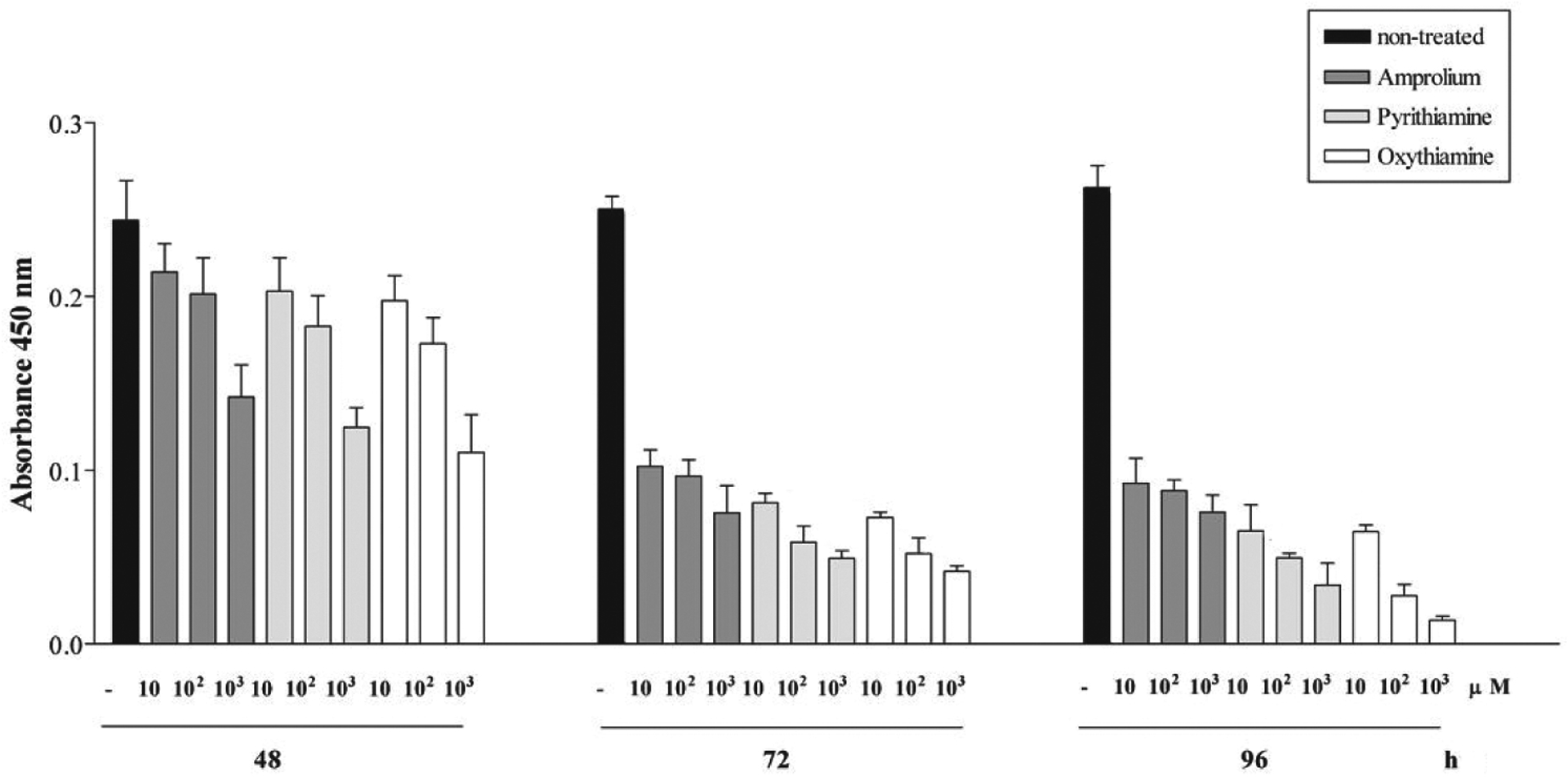
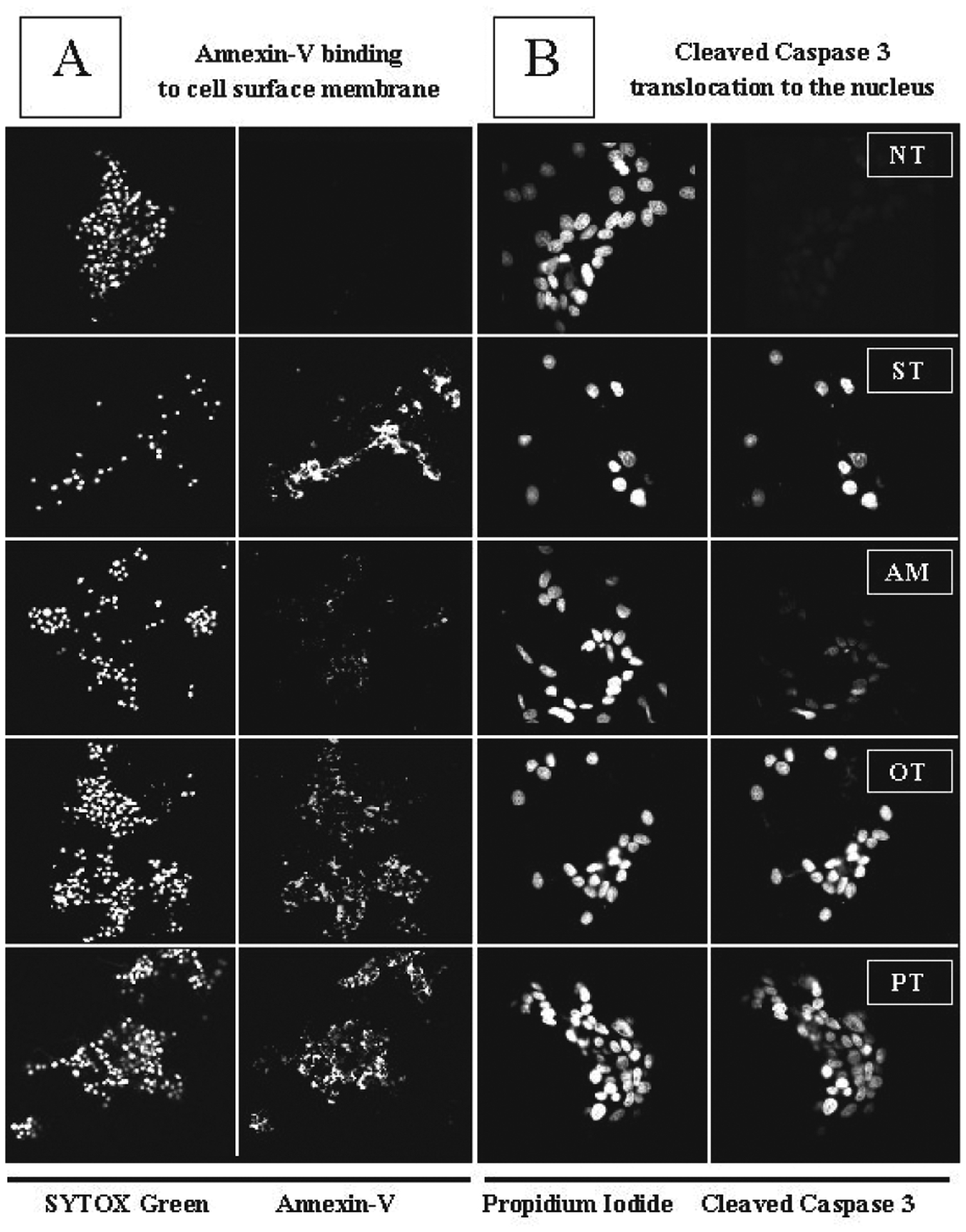
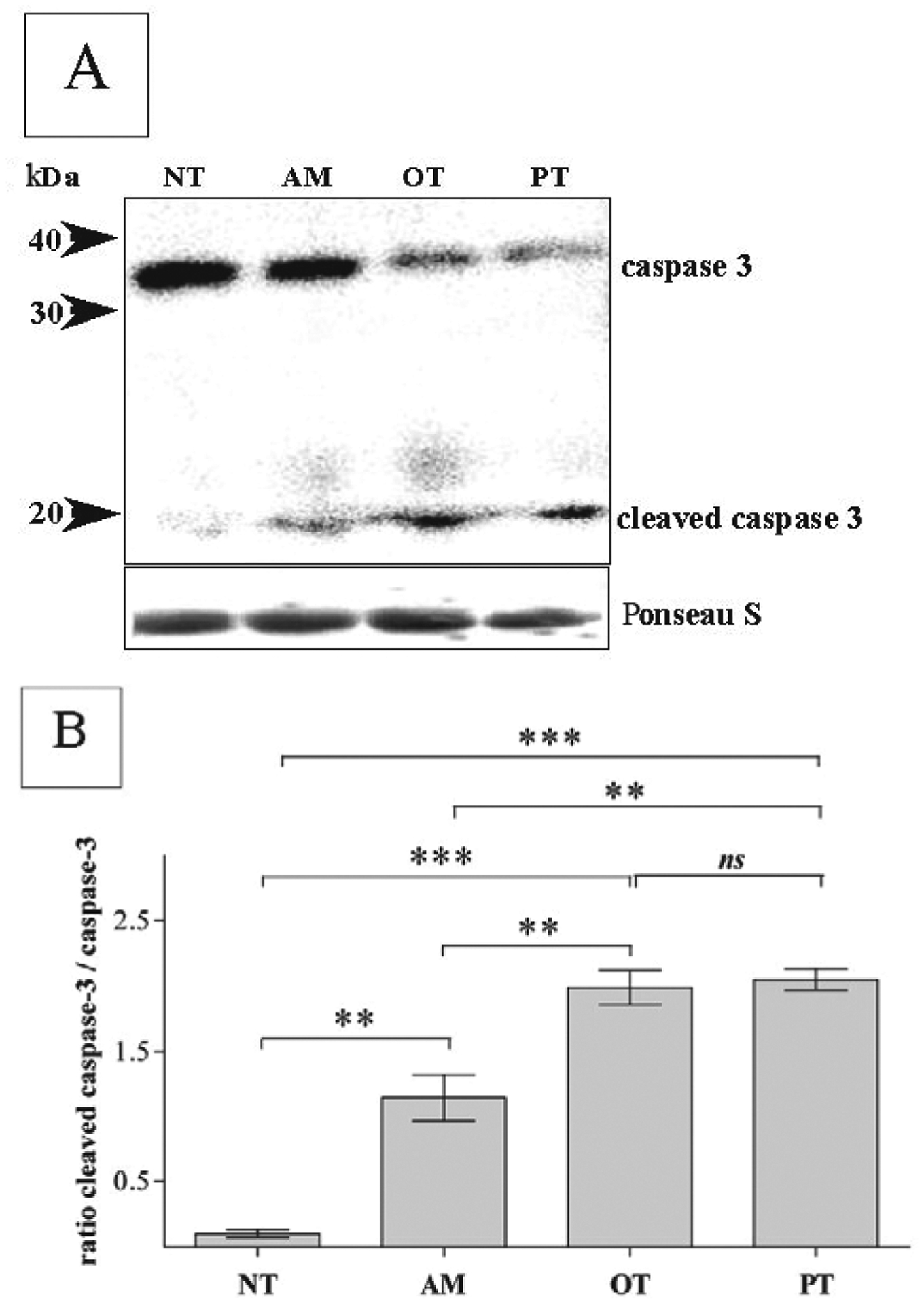
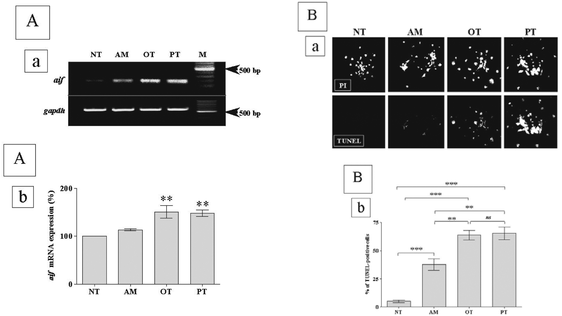
Recent evidence suggests that alterations in oxidative metabolism induced by thiamine deficiency lead to neuronal cell death. However, the molecular mechanisms underlying this process are still under extensive investigation. Here, we report that rat pheochromocytoma PC-12 cells differentiated in the presence of NGF into neurons undergo apoptosis due to thiamine deficiency caused by antagonists of thiamine - amprolium, pyrithiamine and oxythiamine. Confocal laser scanning fluorescence microscopy revealed that annexin V binds to PC-12 cells in presence of thiamine antagonists after 72 h incubation. Results also show that thiamine antagonists trigger upregulation of gene expression of mitochondrial-derived apoptosis inducing factor, DNA fragmentation, cleavage of caspase 3 and translocation of active product to the nucleus. We therefore propose that apoptosis induced by amprolium, pyrithiamine or oxythiamine occurs via the mitochondria-dependent caspase 3-mediated signaling pathway. In addition, our data indicate that pyrithiamine and oxythiamine are more potent inducers of apoptosis than amprolium.
Figures
Similar articles
-
Thiamine and selected thiamine antivitamins - biological activity and methods of synthesis.Biosci Rep. 2018 Jan 10;38(1):BSR20171148. doi: 10.1042/BSR20171148. Print 2018 Feb 28. Biosci Rep. 2018. PMID: 29208764 Free PMC article. Review.
-
Metabolic and structural role of thiamine in nervous tissues.Cell Mol Neurobiol. 2008 Nov;28(7):923-31. doi: 10.1007/s10571-008-9297-7. Epub 2008 Jul 19. Cell Mol Neurobiol. 2008. PMID: 18642074 Free PMC article. Review.
-
Comparative characteristic of thiamine antagonists on apoptosis induction in different types of nerve cell lines.Ukr Biokhim Zh (1999). 2008 Sep-Oct;80(5):76-84. Ukr Biokhim Zh (1999). 2008. PMID: 19248620
-
Thiamine antagonists trigger p53-dependent apoptosis in differentiated SH-SY5Y cells.Sci Rep. 2017 Sep 6;7(1):10632. doi: 10.1038/s41598-017-10878-x. Sci Rep. 2017. PMID: 28878400 Free PMC article.
-
Effect of thiamine deficiency, pyrithiamine and oxythiamine on pyruvate metabolism in rat liver and brain in vivo.J Nutr Sci Vitaminol (Tokyo). 1977;23(5):385-93. doi: 10.3177/jnsv.23.385. J Nutr Sci Vitaminol (Tokyo). 1977. PMID: 604430
Cited by
-
PKR downregulation prevents neurodegeneration and β-amyloid production in a thiamine-deficient model.Cell Death Dis. 2015 Jan 15;6(1):e1594. doi: 10.1038/cddis.2014.552. Cell Death Dis. 2015. PMID: 25590804 Free PMC article.
-
Thiamine and selected thiamine antivitamins - biological activity and methods of synthesis.Biosci Rep. 2018 Jan 10;38(1):BSR20171148. doi: 10.1042/BSR20171148. Print 2018 Feb 28. Biosci Rep. 2018. PMID: 29208764 Free PMC article. Review.
-
Stem Cell Therapy and Thiamine Deficiency-Induced Brain Damage.Neurochem Res. 2024 Jun;49(6):1450-1467. doi: 10.1007/s11064-024-04137-5. Epub 2024 May 9. Neurochem Res. 2024. PMID: 38720090 Review.
-
Metabolic and structural role of thiamine in nervous tissues.Cell Mol Neurobiol. 2008 Nov;28(7):923-31. doi: 10.1007/s10571-008-9297-7. Epub 2008 Jul 19. Cell Mol Neurobiol. 2008. PMID: 18642074 Free PMC article. Review.
-
Amprolium exposure alters mice behavior and metabolism in vivo.Animal Model Exp Med. 2018 Nov 21;1(4):272-281. doi: 10.1002/ame2.12040. eCollection 2018 Dec. Animal Model Exp Med. 2018. PMID: 30891577 Free PMC article.
References
-
- Bakker SJ, Yin M, Kootstra G (1996) Tissue thiamine and carnitine deficiency as a possible cause of acute tubular necrosis after renal transplantation. Transplant Proc 28: 314–315. - PubMed
-
- Bettendorff L, Sluse F, Goessens G, Wins P, Grisar T (1995) Thiamine deficiency-induced partial necrosis and mitochondrial uncoupling in neuroblastoma cells are rapidly reversed by addition of thiamine. J Neurochem 65: 2178–2184. - PubMed
-
- Bradford MM (1976) A rapid and sensitive method for the quantitation of microgram quantities of protein utilizing the principle of protein-dye binding. Anal Biochem 72: 248–254. - PubMed
-
- Butterworth RF, Heroux M (1989) Effects of thiamine deficiency on brain metabolism: implications for the pathogenesis of the Wernicke-Korsakoff syndrome. Alcohol Alcohol 24: 271–279. - PubMed
MeSH terms
Substances
Grants and funding
LinkOut - more resources
Full Text Sources
Research Materials