. 2007 Dec;56(12):1897-910.
doi: 10.1007/s00262-007-0332-1. Epub 2007 May 15.

Intratumoral delivery of vector mediated IL-2 in combination with vaccine results in enhanced T cell avidity and anti-tumor activity

Affiliations

Intratumoral delivery of vector mediated IL-2 in combination with vaccine results in enhanced T cell avidity and anti-tumor activity

Chie Kudo-Saito et al. Cancer Immunol Immunother. 2007 Dec.

Abstract

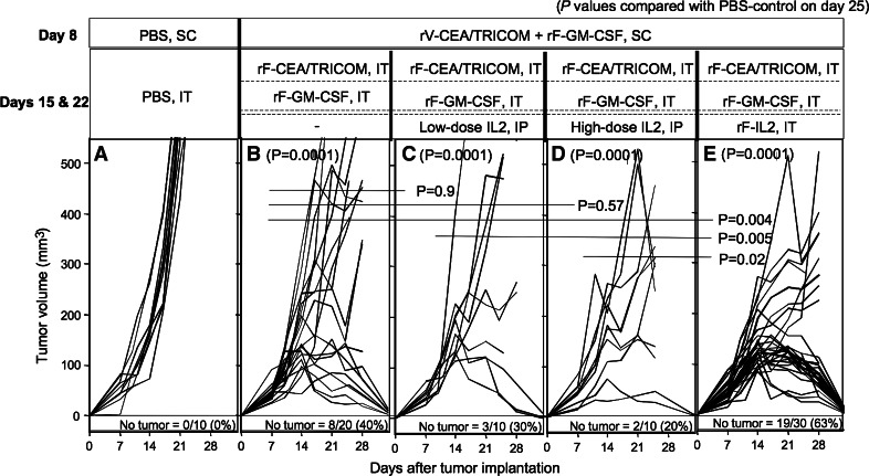
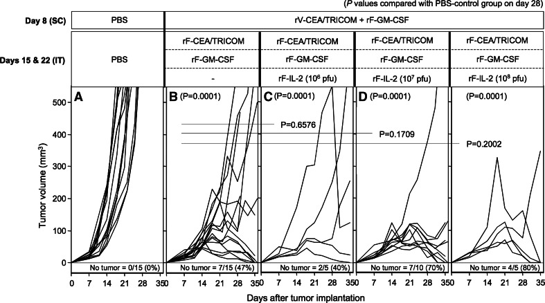
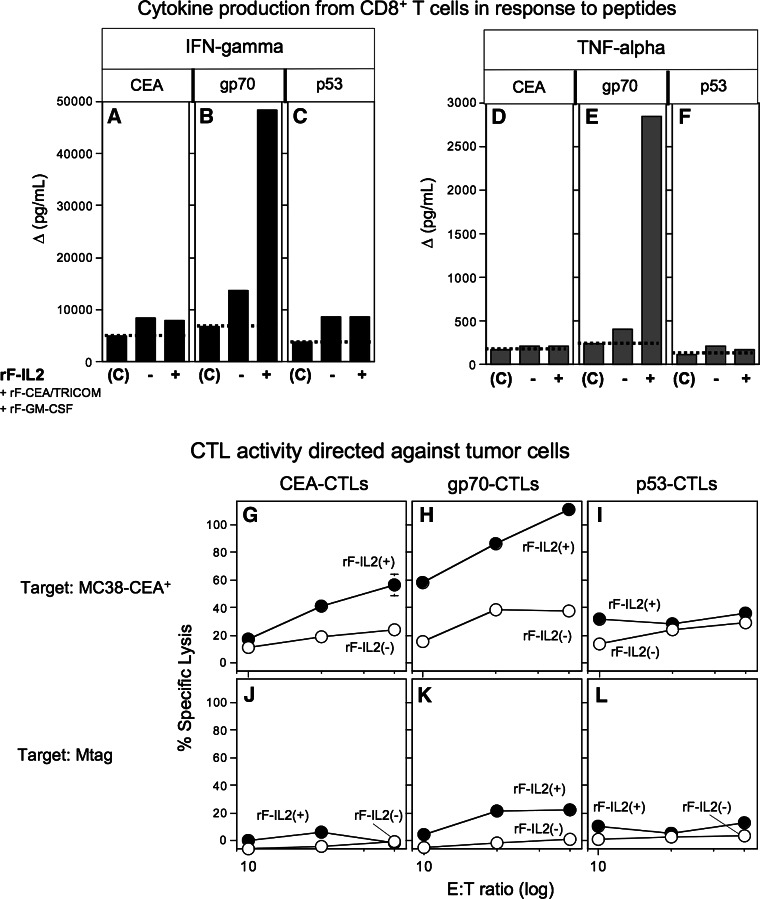
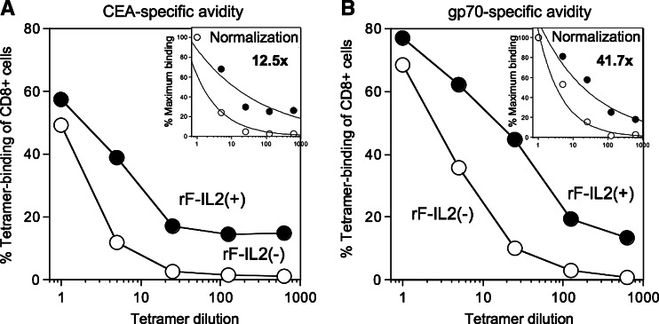
Systemic IL-2 is currently employed in the therapy of several tumor types, but at the price of often severe toxicities. Local vector mediated delivery of IL-2 at the tumor site may enhance local effector cell activity while reducing toxicity. To examine this, a model using CEA-transgenic mice bearing established CEA expressing tumors was employed. The vaccine regimen was a s.c. prime vaccination with recombinant vaccinia (rV) expressing transgenes for CEA and a triad of costimulatory molecules (TRICOM) followed by i.t. boosting with rF-CEA/TRICOM. The addition of intratumoral (i.t.) delivery of IL-2 via a recombinant fowlpox (rF) IL-2 vector greatly enhanced anti-tumor activity of a recombinant vaccine, resulting in complete tumor regression in 70-80% of mice. The anti-tumor activity was shown to be dependent on CD8(+) cells and NK1.1(+). Cellular immune assays revealed that the addition of rF-IL-2 to the vaccination therapy enhanced CEA-specific tetramer(+) cell numbers, cytokine release and CTL lysis of CEA(+) targets. Moreover, tumor-bearing mice vaccinated with the CEA/TRICOM displayed an antigen cascade, i.e., CD8(+) T cell responses to two other antigens expressed on the tumor and not the vaccine: wild-type p53 and endogenous retroviral antigen gp70. Mice receiving rF-IL-2 during vaccination demonstrated higher avidity CEA-specific, as well as higher avidity gp70-specific, CD8(+) T cells when compared with mice vaccinated without rF-IL-2. These studies demonstrate for the first time that the level and avidity of antigen specific CTL, as well as the therapeutic outcome can be improved with the use of i.t. rF-IL-2 with vaccine regimens.


Figures

Fig. 1
Fig. 1
Serum levels of IL-2 after delivery of recombinant IL-2 protein or rF-IL-2. For recombinant protein IL-2 administration, CEA-Tg mice were given low-dose recombinant IL-2 (16,000 IU/dose) i.p. twice a day for four consecutive days (closed triangles). Another group of mice received high-dose recombinant IL-2 (300,000 IU/dose) i.p. twice a day for four consecutive days (closed squares). To measure serum levels of IL-2 following rF-IL-2 administration, mice were injected once with rF-IL-2 (open squares) (107 pfu) s.c. Blood samples were taken at indicated intervals and measured for serum IL-2
Fig. 2
Fig. 2
Systemic IL-2 therapy versus local rF-IL-2 therapy during s.c./i.t. vaccination therapy. CEA-Tg mice were implanted s.c. with MC38-CEA+ tumors on day 0. Mice were vaccinated s.c. on day 8 as a priming vaccination and boosted i.t. on days 15 and 22. a Control mice were administered PBS s.c. on day 8 and i.t. on days 15 and 22. b Mice were vaccinated s.c. with rV-CEA/TRICOM on day 8 and boosted i.t. with rF-CEA/TRICOM on days 15 and 22. c Mice were vaccinated s.c. with rV-CEA/TRICOM on day 8 and boosted i.t. with rF-CEA/TRICOM. Beginning on days 15 and 22, low-dose recombinant IL-2 (16,000 IU/dose) was administered twice a day for four consecutive days. d Mice were vaccinated s.c. with rV-CEA/TRICOM on day 8 and boosted i.t. with rF-CEA/TRICOM. Beginning on days 15 and 22, high-dose recombinant IL-2 (300,000 IU/dose) was administered twice a day for two consecutive days. e Mice were vaccinated s.c. with rV-CEA/TRICOM on day 8 and boosted i.t. with rF-CEA/TRICOM admixed with rF-IL-2 (107 pfu) on days 15 and 22. In groups be, each vaccination was admixed with rF-GM-CSF. Tumor volume was measured one to two times a week. These data are a compilation of three separate experiments
Fig. 3
Fig. 3
Therapeutic efficacy of local doses of rF-IL-2 in combination with s.c./i.t. vaccination using rV-CEA/TRICOM. CEA-Tg mice were implanted s.c. with MC38-CEA+ tumors on day 0. Mice were vaccinated s.c. on day 8 as a priming vaccination and boosted i.t. on days 15 and 22. a Control mice were administered PBS s.c. on day 8 and i.t. on days 15 and 22. b Mice were vaccinated s.c. with rV-CEA/TRICOM on day 8 and boosted i.t. with rF-CEA/TRICOM on days 15 and 22. c Mice were vaccinated s.c. with rV-CEA/TRICOM on day 8 and boosted i.t. with rF-CEA/TRICOM admixed with 106 pfu of rF-IL-2 on days 15 and 22. d Mice were vaccinated s.c. with rV-CEA/TRICOM on day 8 and boosted i.t. with rF-CEA/TRICOM admixed with 107 pfu of rF-IL-2 on days 15 and 22. e Mice were vaccinated s.c. with rV-CEA/TRICOM on day 8 and boosted i.t. with rF-CEA/TRICOM admixed with 108 pfu of rF-IL-2 on days 15 and 22. In groups be, each vaccination was admixed with rF-GM-CSF. Tumor volume was measured one to two times a week. These data are a compilation of two separate experiments
Fig. 4
Fig. 4
Immune cell depletion in mice receiving s.c./i.t. plus local IL-2 vaccination therapy. CEA-transgenic mice were implanted s.c. with MC38-CEA+ tumors on day 0. a Control mice were administered PBS s.c. on day 8, and i.t. on days 15 and 22. be Mice were vaccinated s.c. with rV-CEA/TRICOM on day 8, and boosted i.t. with rF-CEA/TRICOM admixed with rF-IL-2 on days 15 and 22. Each virus was admixed with rF-GM-CSF. b Mice were treated i.p. with PBS during the vaccination therapy. c Mice were treated i.p. with anti-CD4 antibody during the vaccination therapy for CD4+ T cell depletion. d Mice were treated i.p. with anti-CD8 antibody during the vaccination therapy for CD8+ T cell depletion. e Mice were treated i.p. with anti-NK antibody during the vaccination therapy for NK cell depletion. For immune cell depletions, antibodies were administered weekly beginning on days 6, 7 and 8 as described in the “Materials and methods”. Tumor volume was measured one to two times a week
Fig. 5
Fig. 5
Amplification of CD8+ T cell immune responses by rF-IL-2 addition to s.c./i.t. vaccination regimen with rF-CEA/TRICOM. CEA-transgenic mice were implanted s.c. with MC38-CEA+ tumors on day 0. Control mice were administered PBS s.c. on day 8, and i.t. on days 15 and 22 [depicted as (C) in af]. Dotted line represents cytokine level detected in control mice. Mice were vaccinated s.c. with rV-CEA/TRICOM on day 8, and boosted i.t. with rF-CEA/TRICOM with/without (+/−) rF-IL-2 on days 15 and 22. Each virus was admixed with rF-GM-CSF. On day 29 mice were sacrificed and splenic lymphocytes were used for assays (n = 3, pooled). a, d, g and j, splenic lymphocytes were stimulated with CEA class I peptide for 6 days before being tested in assays. b, e, h and k, splenic lymphocytes were stimulated with gp70 class I peptide for 6 days before being tested in assays. c, f, i and l, splenic lymphocytes were stimulated with p53 class I peptide for 6 days before being tested in assays. ac IFN-γ production from CD8+ T cells in response to tumor-antigen class I peptide. Data are depicted as Δ pg/ml; IFN-γ production in response to the control peptide was subtracted from that induced by tumor-antigen peptide. a CEA-specific IFN-γ production. b gp70-specific IFN-γ production. c p53-specific IFN-γ production. df TNF-α production from CD8+ T cells in response to tumor-antigen class I peptide. Data are depicted as Δ pg/ml; TNF-α production in response to the control peptide was subtracted from that induced by tumor-antigen peptide. d CEA-specific TNF-α production. e gp70-specific TNF-α production. f p53-specific TNF-α production. gi CTL activity against MC38-CEA+ tumor cells. jl CTL activity against Mtag tumor cells. SD based on the mean of triplicate wells. Open circles, mice boosted i.t. with rF-CEA/TRICOM (without rF-IL-2) after prime vaccination with rV-CEA/TRICOM. Closed circles, mice boosted i.t. with rF-CEA/TRICOM admixed with rF-IL-2 after prime vaccination with rV-CEA/TRICOM. These data are representative of three independent experiments
Fig. 6
Fig. 6
Induction of tumor-specific CD8+ T cells in tumor sites in mice receiving CEA/TRICOM s.c./i.t. vaccination therapy. CEA-transgenic mice were implanted s.c. with MC38-CEA+ tumors on day 0. Mice were vaccinated s.c. with rV-CEA/TRICOM on day 8, and boosted i.t. on days 15 and 22 with the virus indicated. Each virus was admixed with rF-GM-CSF (GM). These mice were sacrificed and TILs were used for assays 7 days after the last i.t. boost vaccination (day 29 after tumor implantation, n = 3, pooled). ac CEA-specific CD8+ T cells. Cells were stained with anti-CD3 mAb, anti-CD8 mAb and CEA-tetramer. df gp70-specific CD8+ T cells. Cells were stained with anti-CD3 mAb, anti-CD8 mAb and gp70-tetramer. Stained cells were first gated on the CD3+CD8+ cell fraction. The numbers indicate the percentage of tetramer+ cells of CD3+CD8+ cells. These data are representative of three independent experiments
Fig. 7
Fig. 7
Enhancement of T cell avidity of CD8+ cells by rF-IL-2 addition to s.c./i.t. vaccination regimen. CEA-transgenic mice were implanted s.c. with MC38-CEA+ tumors on day 0. Mice were vaccinated s.c. with rV-CEA/TRICOM on day 8, and boosted i.t. with rF-CEA/TRICOM with/without (+/−) rF-IL-2 on days 15 and 22. Each virus was admixed with rF-GM-CSF. Mice were sacrificed, and TILs were used for assays 7 days after the last i.t. boost vaccination (day 29 after tumor implantation, n = 3, pooled). a CEA-specific CD8+ T cell avidity as determined by dilutional tetramer staining. TILs were stained with anti-CD3 mAb, anti-CD8 mAb and diluted CEA-tetramer (1×, 5×, 25×, 125×, 625× dilution). The results were depicted as the percentage of CEA-tetramer+ cells of CD8+ T cells. The inset shows the results depicted as the percentage of maximum tetramer-binding cells to normalize groups within each experiment. The percent of tetramer-binding cells detected without dilution of tetramer (1 μl/106 cells) was calculated as 100%. Tetramer binding efficacy was determined by calculating the reciprocal of the tetramer dilution that resulted in 50% of the normalized maximum tetramer binding. Fold change was derived mathematically from the difference between the individual group values. b gp70-specific CD8+ cell avidity as determined by dilutional tetramer staining. TILs were stained with anti-CD3 mAb, anti-CD8 mAb and diluted gp70-tetramer (1×, 5×, 25×, 125×, 625× dilution). The results were depicted as the percentage of gp70-tetramer+ cells of CD8+ cells. The inset shows the results depicted as the percentage of maximum tetramer-binding to normalize groups within each experiment. Open circles, mice boosted i.t. with rF-CEA/TRICOM (without rF-IL-2). Closed circles, mice boosted i.t. with rF-CEA/TRICOM admixed with rF-IL-2. These data are representative of two independent experiments

Similar articles

Cited by

References

    1. Aarts WM, Schlom J, Hodge JW. Vector-based vaccine/cytokine combination therapy to enhance induction of immune responses to a self-antigen and antitumor activity. Cancer Res. 2002;62:5770. - PubMed
    1. Chakraborty M, Abrams SI, Coleman CN, Camphausen K, Schlom J, Hodge JW. External beam radiation of tumors alters phenotype of tumor cells to render them susceptible to vaccine-mediated T-cell killing. Cancer Res. 2004;64:4328. doi: 10.1158/0008-5472.CAN-04-0073. - DOI - PubMed
    1. Chen B, Timiryasova TM, Andres ML, Kajioka EH, Dutta-Roy R, Gridley DS, Fodor I. Evaluation of combined vaccinia virus-mediated antitumor gene therapy with p53, IL-2, and IL-12 in a glioma model. Cancer Gene Ther. 2000;7:1437. doi: 10.1038/sj.cgt.7700252. - DOI - PubMed
    1. Dillman RO. What to do with IL-2? Cancer Biother Radiopharm. 1999;14:423. doi: 10.1089/cbr.1999.14.423. - DOI - PubMed
    1. Disis ML, Bernhard H, Shiota FM, Hand SL, Gralow JR, Huseby ES, Gillis S, Cheever MA. Granulocyte-macrophage colony-stimulating factor: an effective adjuvant for protein and peptide-based vaccines. Blood. 1996;88:202. - PubMed

Publication types