

Membrane organization of the serotonin 1A receptor monitored by a detergent-free approach
- PMID: 17503188
- PMCID: PMC11881808
- DOI: 10.1007/s10571-007-9138-0
Membrane organization of the serotonin 1A receptor monitored by a detergent-free approach
Abstract
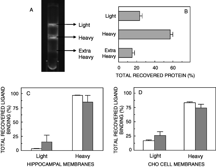
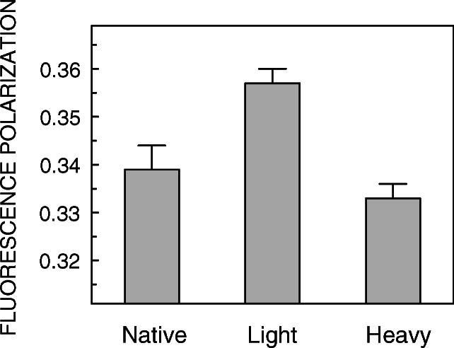
: 1. Insolubility of membrane constituents in nonionic detergents such as Triton X-100 has been a widely used biochemical criterion to indicate their localization in membrane domains. However, concerns on the possibility of membrane perturbation in the presence of detergents have led to the development of detergent-free approaches.2. We have explored the organization of the serotonin(1A) receptor, an important G-protein coupled receptor, from bovine hippocampus and CHO cells using a detergent-free approach in order to address the points of agreement with our previous results using Triton X-100.3. A significant fraction of the serotonin(1A) receptor has been found to be localized in a heavy density fraction obtained using a detergent-free approach to isolate membrane domains. In addition, we have characterized the membrane fractions isolated in terms of their lipid composition and membrane physical properties.4. The results obtained on the membrane localization of the serotonin(1A) receptor from the present experiments using a detergent-free approach correlate well with our earlier findings obtained using a detergent-based method (Kalipatnapu, S., and Chattopadhyay, A., FEBS Lett. 576:455-460, 2004). These results provide important information on the membrane organization of the hippocampal serotonin(1A) receptor and are relevant in view of the concerns on the use of detergent in determination of membrane organization of constituent proteins and lipids.
Figures
Similar articles
-
Membrane organization and function of the serotonin(1A) receptor.Cell Mol Neurobiol. 2007 Dec;27(8):1097-116. doi: 10.1007/s10571-007-9189-2. Epub 2007 Aug 21. Cell Mol Neurobiol. 2007. PMID: 17710529 Free PMC article. Review.
-
Cholesterol depletion modulates detergent resistant fraction of human serotonin(1A) receptors.Mol Membr Biol. 2012 Nov;29(7):290-8. doi: 10.3109/09687688.2012.688147. Epub 2012 May 18. Mol Membr Biol. 2012. PMID: 22594670
-
Membrane organization of the human serotonin(1A) receptor monitored by detergent insolubility using GFP fluorescence.Mol Membr Biol. 2005 Nov-Dec;22(6):539-47. doi: 10.1080/09687860500421738. Mol Membr Biol. 2005. PMID: 16373325
-
A GFP fluorescence-based approach to determine detergent insolubility of the human serotonin1A receptor.FEBS Lett. 2004 Oct 22;576(3):455-60. doi: 10.1016/j.febslet.2004.09.055. FEBS Lett. 2004. PMID: 15498580
-
Membrane organization and dynamics of the serotonin1A receptor in live cells.J Neurochem. 2011 Mar;116(5):726-33. doi: 10.1111/j.1471-4159.2010.07037.x. Epub 2011 Jan 7. J Neurochem. 2011. PMID: 21214564 Review.
Cited by
-
Membrane organization and function of the serotonin(1A) receptor.Cell Mol Neurobiol. 2007 Dec;27(8):1097-116. doi: 10.1007/s10571-007-9189-2. Epub 2007 Aug 21. Cell Mol Neurobiol. 2007. PMID: 17710529 Free PMC article. Review.
-
Designing therapies against experimental visceral leishmaniasis by modulating the membrane fluidity of antigen-presenting cells.Infect Immun. 2009 Jun;77(6):2330-42. doi: 10.1128/IAI.00057-09. Epub 2009 Mar 16. Infect Immun. 2009. PMID: 19289510 Free PMC article.
References
-
- Amundson, D. M., and Zhou, M. (1999). Fluorometric method for the enzymatic determination of cholesterol. J. Biochem. Biophys. Methods38:43–52. - PubMed
-
- Blier, P., de Montigny, C., and Chaput, Y. (1990). A role for the serotonin system in the mechanism of action of antidepressant treatments: Preclinical evidence. J. Clin. Psychiatry51:14–20. - PubMed
-
- Bligh, E. G., and Dyer, W. J. (1959). A rapid method of total lipid extraction and purification. Can. J. Biochem. Physiol.37:911–917. - PubMed
-
- Brown, D. A., and London, E. (1998). Structure and origin of ordered lipid domains in biological membranes. J. Membr. Biol.164:103–114. - PubMed
-
- Brown, D. A., and Rose, J. K. (1992). Sorting of GPI-anchored proteins to glycolipid-enriched membrane subdomains during transport to the apical cell surface. Cell68:533–544. - PubMed
Publication types
MeSH terms
Substances
LinkOut - more resources
Full Text Sources
