Review
. 2007 Nov;20(7):633-42.
doi: 10.1002/nbm.1177.

Measurement of cerebral perfusion territories using arterial spin labelling

Affiliations
Review

Measurement of cerebral perfusion territories using arterial spin labelling

Fernando F Paiva et al. NMR Biomed. 2007 Nov.

Abstract

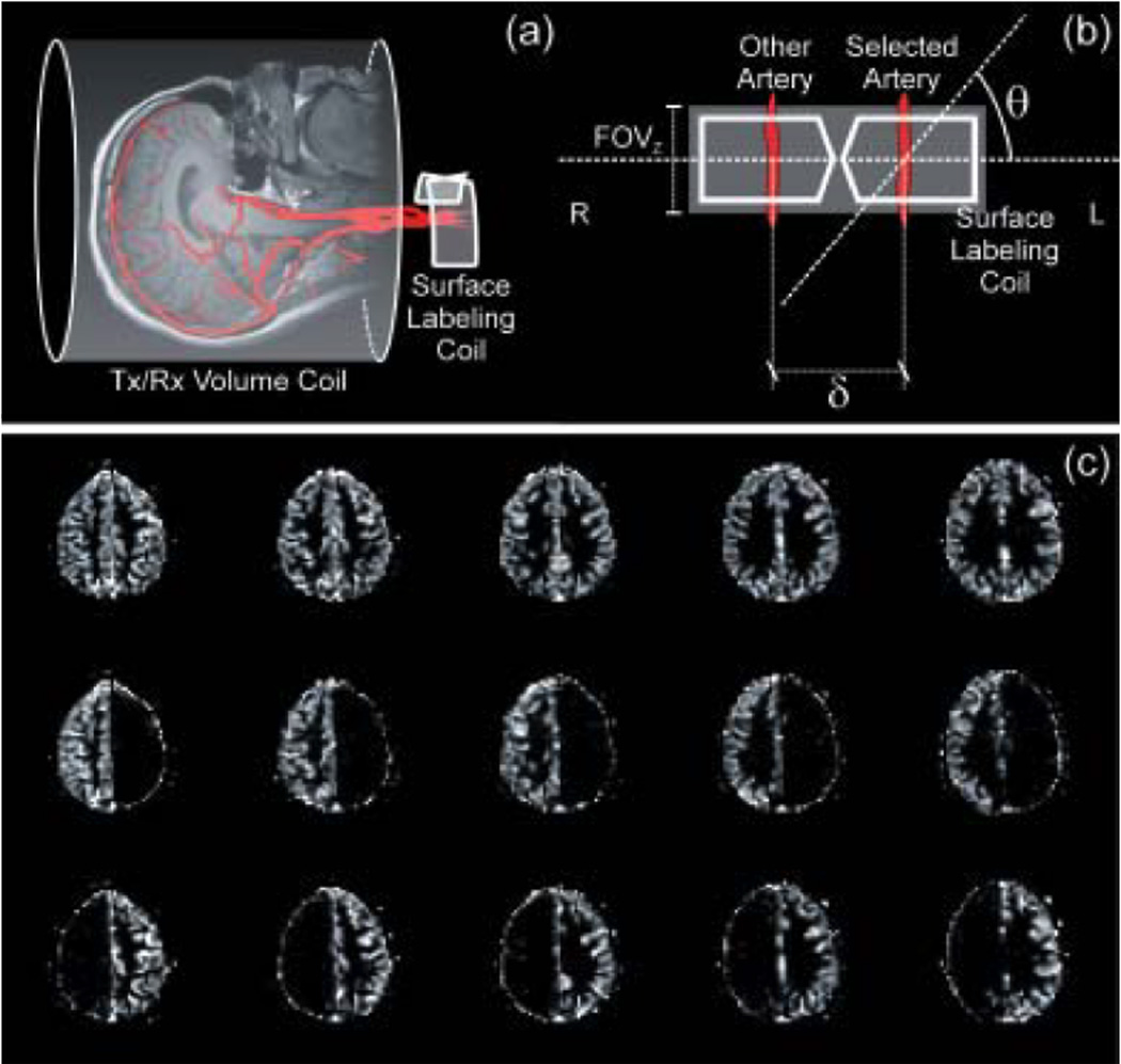
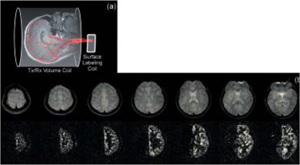
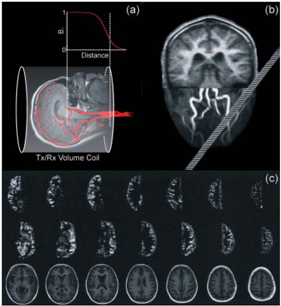
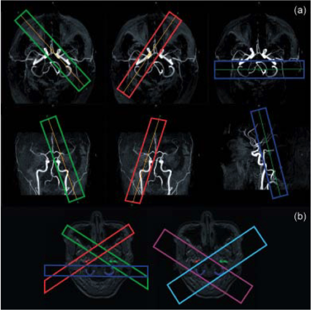
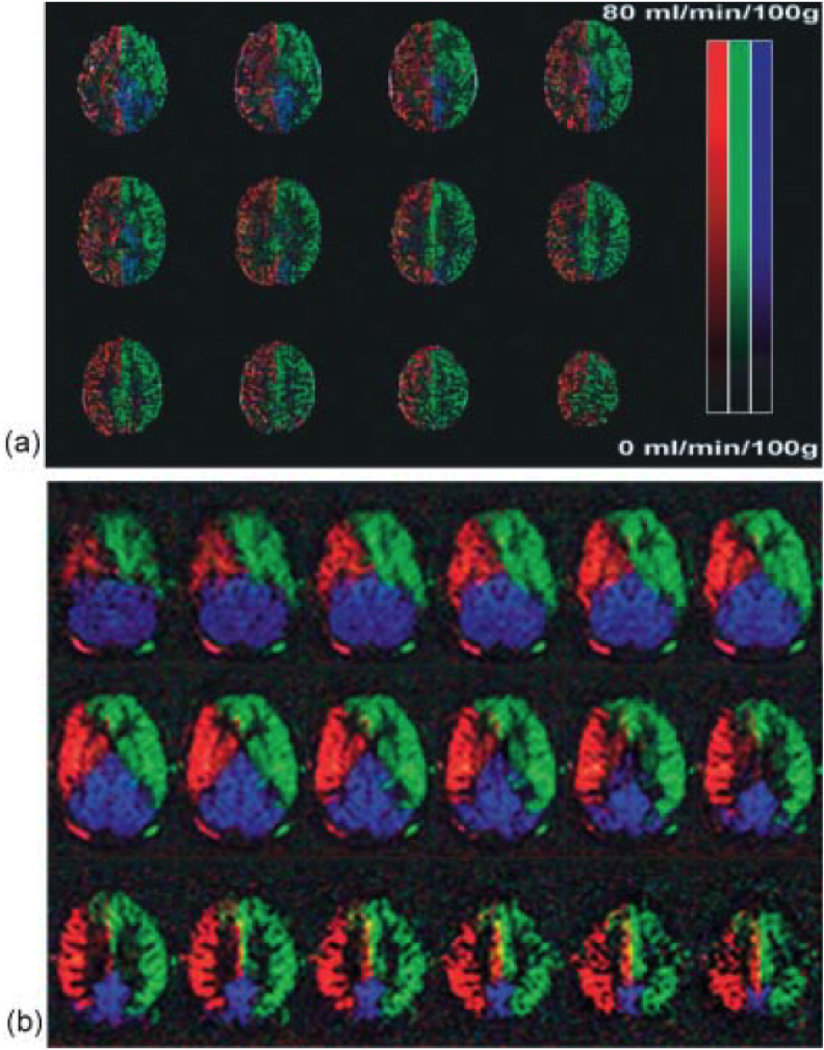
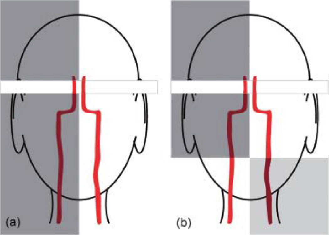
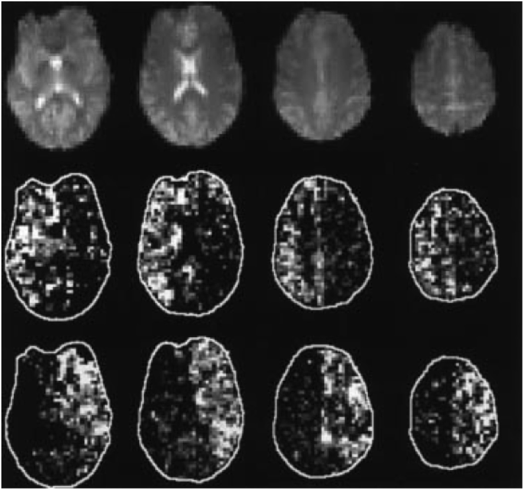
The ability to assess the perfusion territories of major cerebral arteries can be a valuable asset to the diagnosis of a number of cerebrovascular diseases. Recently, several arterial spin labeling (ASL) techniques have been proposed for determining the cerebral perfusion territories of individual arteries by three different approaches: (1) using a dedicated labeling radio frequency (RF) coil; (2) applying selective inversion of spatially confined areas; (3) employing multidimensional RF pulses. Methods that use a separate labeling RF coil have high signal-to-noise ratio (SNR), low RF power deposition, and unrestricted three-dimensional coverage, but are mostly limited to separation of the left and right circulation, and do require extra hardware, which may limit their implementation in clinical systems. Alternatively, methods that utilize selective inversion have higher flexibility of implementation and higher arterial selectivity, but suffer from imaging artifacts resulting from interference between the labeling slab and the volume of interest. The goal of this review is to provide the reader with a critical survey of the different ASL approaches proposed to date for determining cerebral perfusion territories, by discussing the relative advantages and disadvantages of each technique, so as to serve as a guide for future refinement of this promising methodology.


Figures

Figure 1
Figure 1
(a) Schematic representation of the separate labeling/imaging coil approach. The labeling RF coil is placed right over the neck to access both CCAs and VAs. (b) An oblique labeling plane, employed at an angle θ with respect to a plane perpendicular to blood flow, allows for selective labeling of the desired arteries. Positive values of θ target the left circulation, while negative values target the right circulation. (c) CBF images of a healthy human subject, obtained by setting θ = 0° (top row), θ = −60° (middle row) and θ = +60° (bottom), corresponding to the CBF images of the whole brain, the right and the left circulation, respectively.
Figure 2
Figure 2
(a) An alternative option that excludes the need of the oblique labeling plane is to place the labeling RF coil right over one of the carotids. (b) Multi-slice axial control (top) and subtraction (bottom) images created using a unilateral carotid labeling RF coil and spin echo EPI (TR/TE/labeling period/post-labeling delay = 4s/22ms/3s/0.5s). As expected in the normal brain, perfusion from one carotid artery supplies only the ipsilateral hemisphere. The excellent subtraction of the contralateral hemisphere serves as a demonstration of the elimination of magnetization transfer effects. This method requires repositioning the coil to selectively label the contra lateral carotid. (Reproduced by permission of Zaharchuk et al (15)).
Figure 3
Figure 3
(a) Schematic illustration of the amplitude profile along the longitudinal axis of a standard transmitter/receive volume coil. The selective label of the desired arteries is achieved exploring the limited longitudinal field of the volume coil in the neck’s region combined with an oblique labeling plane. (b) Positioning of the labeling plane during the MR experiment based on maximum intensity projection of angiography data. The image of the brain was added for illustration purposes. The oblique plane intersects the selected arteries 80 mm, the contralateral arteries 160 mm below the coil center. Adapted from Werner et al (20). Perfusion-weighted images as acquired in a volunteer. First/second row: perfusion images with left/right-sided arteries labeled; bottom row: anatomical images. The oblique-plane selection mechanism yields clearly delineated perfusion territory images of the anterior circulation. Gray and white matter can easily be distinguished even in the smaller structures. (Reproduced by permission of Werner et al (20)).
Figure 4
Figure 4
(a) Planning of the respective labeling of the left ICA (green), right ICA (red), and posterior circulation (blue) on the MIPs of the circle of Willis of a healthy volunteer. (Reproduced by permission of Golay et al (25)) (b) Planning of the labeling scheme used in the dual vessel approach, in which two acquisitions (light blue and purple), both including the posterior circulation, replace the three acquisitions (red, green and blue) from the previous method. (Courtesy of Dr. Xavier Golay).
Figure 5
Figure 5
Color-coded based territories maps of the left ICA (green), right ICA (red), and posterior circulation (blue) from healthy volunteers. (a) Maps acquired based on three independent acquisitions. (Reproduced by permission of Golay et al (25)). (b) Maps acquired in a single cycled ASL experiment and processed using independent component analysis. (Reproduced by permission of Günther (27)).
Figure 6
Figure 6
Schematic representation of selectively labeling schemes that combine RF pulses and saturation of the imaging slices to achieve better definition of the vascular territory. (a) A sagittal unilateral selective inversion pulse inverts the spins in the selected carotid. A saturation pulse followed by a spoiler gradient is applied to destroy the effects of the inversion pulse in the imaging slice. (b) A sagittal unilateral selective inversion pulse is followed by a transverse selective inversion pulse that swaps the labeling scheme in the region located below the acquisition slice. A saturation pulse followed by a spoiler gradient is applied to destroy the effects of the inversion pulse in the imaging slice.
Figure 7
Figure 7
(a) Schematic illustration of the rotating labeling plane scheme used to selectively label the desired artery (23). A rotating labeling gradient G combined with a frequency-modulated labeling RF pulse sets the position of the labeling plane to the selected artery while the adjacent areas observes a sinusoidally-variable resonant behavior. The shaded area illustrates the moving labeling plane coverage. (b) Perfusion territory images acquired in a health volunteer, obtained with the Continuous Artery-Selective Spin Labeling (CASSL) scheme (23). Top row: T1-weighted anatomical reference images. Second row: right-sided MCA selected. Third row: left-sided MCA selected. Bottom row: left- and right-sided ACAs selected. The perfusion territories are clearly delineated. Regions in which the labeling mechanism intersects the imaging volume appear dark. (Reproduced by permission of Werner et al (23)).
Figure 8
Figure 8
The four superior slices of baseline EPI readout (M0) from one subject (top panel). Also shown are mean pairwise difference images normalize to baseline EPI (M/M0) resulting from a 2D tag applied to the right ICA (middle panel) and the left ICA (bottom panel), selected with the use of a tailored 2-dimensional RF pulse. (Reproduced by permission of Davies and Jezzard (18)).

References

    1. Hoeffner EG. Cerebral perfusion imaging. J Neuroophthalmol. 2005;25(4):313–320. - PubMed
    1. Wintermark M, Sesay M, Barbier E, Borbely K, Dillon WP, Eastwood JD, Glenn TC, Grandin CB, Pedraza S, Soustiel JF, Nariai T, Zaharchuk G, Caille JM, Dousset V, Yonas H. Comparative overview of brain perfusion imaging techniques. Stroke. 2005;36(9):e83–e99. - PubMed
    1. Cha S. Perfusion MR imaging: basic principles and clinical applications. Magn Reson Imaging Clin N Am. 2003;11(3):403–413. - PubMed
    1. Lad SP, Guzman R, Kelly ME, Li G, Lim M, Lovbald K, Steinberg GK. Cerebral perfusion imaging in vasospasm. Neurosurg Focus. 2006;21(3):E7. - PubMed
    1. van Laar PJ, Hendrikse J, Golay X, Lu H, van Osch MJ, van der Grond J. In vivo flow territory mapping of major brain feeding arteries. Neuroimage. 2006;29(1):136–144. - PubMed

Publication types

LinkOut - more resources