

Ischaemia and reperfusion effects on skeletal muscle tissue: morphological and histochemical studies
- PMID: 17504444
- PMCID: PMC2517305
- DOI: 10.1111/j.1365-2613.2007.00526.x
Ischaemia and reperfusion effects on skeletal muscle tissue: morphological and histochemical studies
Abstract
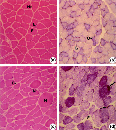
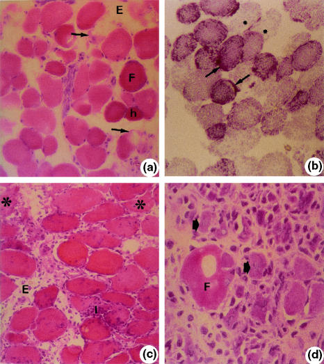
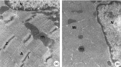
This was a study on the oxidative stress due to ischaemia (I) and reperfusion (R) in skeletal muscle tissue. Using a tourniquet, groups of rats were submitted to ischaemia for 4 h, followed by different reperfusion periods. The animals were divided in four groups: control; 4 h of ischaemia (IR); 4 h of ischaemia plus 1 h reperfusion (IR-1 h); 4 h of ischaemia plus 24 h reperfusion (IR-24 h); and 4 h of ischaemia plus 72 h reperfusion (IR-72 h). At the end of the procedures, samples of soleus muscle were collected and frozen in n-hexane at -70 degrees C. Cryostat sections were submitted to haematoxylin-eosin, succinate dehydrogenase (SDH) and nicotinamide adenine dinucleotide-tetrazolium reductase (NADH-TR) stains. An additional muscle sample was processed for electron microscopy. No alterations were found in control animals. IR group showed fibres had normal aspect besides some round, acidophilic and hypertrophic fibres. There were several fibres with angular outlines and smaller diameters in this group compared with control group. NADH-TR/SDH reaction was moderately intense in most fibres. In some fibres, cytoplasm showed areas without activity and other fibres had very intense reactivity. IR-1 h group showed oedema hypercontracted fibres with disorganized myofibrils, mitochondria with focal lesions and dilated sarcoplasmic reticulum. NADH-TR/SDH reaction was moderate to weak. IR-24 h showed intense inflammatory infiltrate in the endomysium and perimysium. NADH-TR/SDH reaction was similar to IR-1 h. IR-72 h showed necrotic fibres, areas with inflammatory infiltrate, reduced muscle fibres at different stages of necrosis and phagocytosis, and many small round and basophilic fibres characterizing a regeneration process. NADH-TR/SDH reaction was weak to negative. Our results suggest that ischaemia and the subsequent 1-, 24- and 72-h reperfusions induced progressive histological damage. Although progressive, it may be reversible because there were ultrastructural signs of recovery after 72-h reperfusion. This recovery could in part be due to the low oxidative stress identified by the morphological and histochemical analysis.
Figures

References
-
- Appell HJ, Duarte JA, Gloser S, et al. Administration of tourniquet. II. Prevention of postischemic oxidative stress can reduce muscle edema. Arch. Orthop. Trauma Surg. 1997;116:101–105. - PubMed
-
- Beyersdorf F, Matheis G, Kruger S, et al. Avoiding reperfusion injury after limb revascularization: experimental observations and recommendations for clinical application. J. Vasc. Surg. 1989;6:757–766. - PubMed
-
- Bigard AX, Boehm E, Veksler V, Mateo P, Anflous K, Ventura-Clapier R. Muscle unloading induces slow to fast transitions in myofibrillar but not mitochondrial properties. Relevance to skeletal muscle abnormalities in heart failure. J. Mol. Cell. Cardiol. 1998;30:2391–2401. - PubMed
-
- Blaisdell FW, Steele M, Allen RE. Management of acute lower extremity arterial ischemia due to embolism and thrombosis. Surgery. 1978;84:822–834. - PubMed
-
- Bushell AJ, Klenerman L, Davies H, Grierson I, McArdle A, Jackson MJ. Ischaemic preconditioning of skeletal muscle 2. Investigation of the potential mechanisms involved. J. Bone Joint Surg. 2002;84:1189–1193. - PubMed
Publication types
MeSH terms
LinkOut - more resources
Full Text Sources
Other Literature Sources
