Isolated receptor binding domains of HTLV-1 and HTLV-2 envelopes bind Glut-1 on activated CD4+ and CD8+ T cells
- PMID: 17504522
- PMCID: PMC1876471
- DOI: 10.1186/1742-4690-4-31
Isolated receptor binding domains of HTLV-1 and HTLV-2 envelopes bind Glut-1 on activated CD4+ and CD8+ T cells
Abstract
Background: We previously identified the glucose transporter Glut-1, a member of the multimembrane-spanning facilitative nutrient transporter family, as a receptor for both HTLV-1 and HTLV-2. However, a recent report concluded that Glut-1 cannot serve as a receptor for HTLV-1 on CD4 T cells: This was based mainly on their inability to detect Glut-1 on this lymphocyte subset using the commercial antibody mAb1418. It was therefore of significant interest to thoroughly assess Glut-1 expression on CD4 and CD8 T cells, and its association with HTLV-1 and -2 envelope binding.
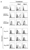
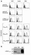
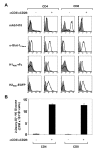
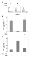
Results: As previously reported, ectopic expression of Glut-1 but not Glut-3 resulted in significantly augmented binding of tagged proteins harboring the receptor binding domains of either HTLV-1 or HTLV-2 envelope glycoproteins (H1RBD or H2RBD). Using antibodies raised against the carboxy-terminal peptide of Glut-1, we found that Glut-1 expression was significantly increased in both CD4 and CD8 cells following TCR stimulation. Corresponding increases in the binding of H1RBD as well as H2RBD, not detected on quiescent T cells, were observed following TCR engagement. Furthermore, increased Glut-1 expression was accompanied by a massive augmentation in glucose uptake in TCR-stimulated CD4 and CD8 lymphocytes. Finally, we determined that the apparent contradictory results obtained by Takenouchi et al were due to their monitoring of Glut-1 with a mAb that does not bind cells expressing endogenous Glut-1, including human erythrocytes that harbor 300,000 copies per cell.
Conclusion: Transfection of Glut-1 directly correlates with the capacities of HTLV-1 and HTLV-2 envelope-derived ligands to bind cells. Moreover, Glut-1 is induced by TCR engagement, resulting in massive increases in glucose uptake and binding of HTLV-1 and -2 envelopes to both CD4 and CD8 T lymphocytes. Therefore, Glut-1 is a primary binding receptor for HTLV-1 and HTLV-2 envelopes on activated CD4 as well as CD8 lymphocytes.
Figures


Similar articles
-
In vivo cellular tropism of human T-lymphotropic virus type II is not restricted to CD8+ cells.Virology. 1995 Jul 10;210(2):441-7. doi: 10.1006/viro.1995.1360. Virology. 1995. PMID: 7542419
-
HTLV-1 and -2 envelope SU subdomains and critical determinants in receptor binding.Retrovirology. 2004 Dec 2;1:41. doi: 10.1186/1742-4690-1-41. Retrovirology. 2004. PMID: 15575958 Free PMC article.
-
Infection of CD4+ T lymphocytes by the human T cell leukemia virus type 1 is mediated by the glucose transporter GLUT-1: evidence using antibodies specific to the receptor's large extracellular domain.Virology. 2006 May 25;349(1):184-96. doi: 10.1016/j.virol.2006.01.045. Epub 2006 Mar 7. Virology. 2006. PMID: 16519917
-
HTLV-1 tropism and envelope receptor.Oncogene. 2005 Sep 5;24(39):6016-25. doi: 10.1038/sj.onc.1208972. Oncogene. 2005. PMID: 16155608 Review.
-
[HTLV-I--associated myelopathy/tropical spastic paraparesis (HAM/TSP)].Ryoikibetsu Shokogun Shirizu. 2000;(31):9-12. Ryoikibetsu Shokogun Shirizu. 2000. PMID: 11269197 Review. Japanese. No abstract available.
Cited by
-
Human T-cell leukemia virus type I infects human lung epithelial cells and induces gene expression of cytokines, chemokines and cell adhesion molecules.Retrovirology. 2008 Sep 22;5:86. doi: 10.1186/1742-4690-5-86. Retrovirology. 2008. PMID: 18808681 Free PMC article.
-
Knockdown of synapse-associated protein Dlg1 reduces syncytium formation induced by human T-cell leukemia virus type 1.Virus Genes. 2008 Aug;37(1):9-15. doi: 10.1007/s11262-008-0234-0. Epub 2008 May 7. Virus Genes. 2008. PMID: 18461433
-
Entry of glucose- and glutamine-derived carbons into the citric acid cycle supports early steps of HIV-1 infection in CD4 T cells.Nat Metab. 2019 Jul;1(7):717-730. doi: 10.1038/s42255-019-0084-1. Epub 2019 Jul 12. Nat Metab. 2019. PMID: 32373781 Free PMC article.
-
Cell surface Glut1 levels distinguish human CD4 and CD8 T lymphocyte subsets with distinct effector functions.Sci Rep. 2016 Apr 12;6:24129. doi: 10.1038/srep24129. Sci Rep. 2016. PMID: 27067254 Free PMC article.
-
Glucose Metabolism in T Cells and Monocytes: New Perspectives in HIV Pathogenesis.EBioMedicine. 2016 Apr;6:31-41. doi: 10.1016/j.ebiom.2016.02.012. Epub 2016 Feb 6. EBioMedicine. 2016. PMID: 27211546 Free PMC article. Review.
References
-
- Kim FJ, Seiliez I, Denesvre C, Lavillette D, Cosset FL, Sitbon M. Definition of an amino-terminal domain of the human T-cell leukemia virus type 1 envelope surface unit that extends the fusogenic range of an ecotropic murine leukemia virus. J Biol Chem. 2000;275:23417–23420. doi: 10.1074/jbc.C901002199. - DOI - PubMed
Publication types
MeSH terms
Substances
LinkOut - more resources
Full Text Sources
Other Literature Sources
Research Materials
Miscellaneous

