. 2007 May 15:4:32.
doi: 10.1186/1742-4690-4-32.

Antigen-presenting particle technology using inactivated surface-engineered viruses: induction of immune responses against infectious agents

Affiliations

Antigen-presenting particle technology using inactivated surface-engineered viruses: induction of immune responses against infectious agents

Joseph D Mosca et al. Retrovirology. .

Abstract

Background: Developments in cell-based and gene-based therapies are emerging as highly promising areas to complement pharmaceuticals, but present day approaches are too cumbersome and thereby limit their clinical usefulness. These shortcomings result in procedures that are too complex and too costly for large-scale applications. To overcome these shortcomings, we described a protein delivery system that incorporates over-expressed proteins into viral particles that are non-infectious and stable at room temperature. The system relies on the biological process of viral egress to incorporate cellular surface proteins while exiting their host cells during lytic and non-lytic infections.

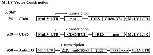
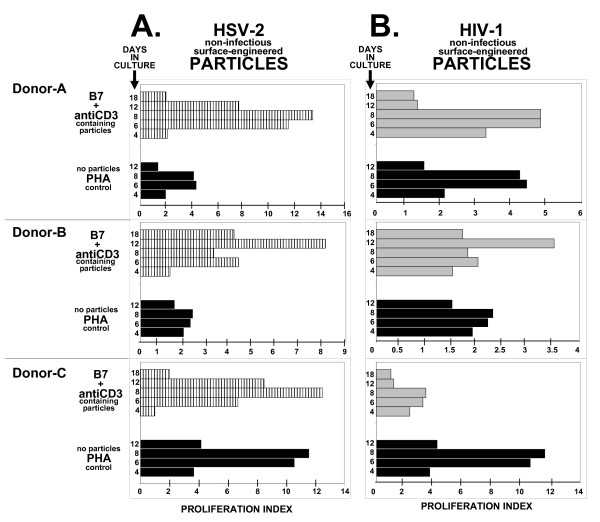
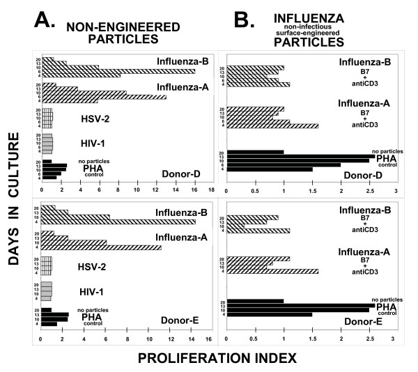
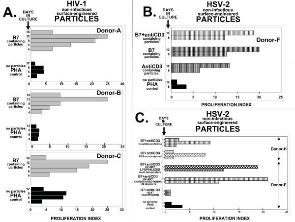
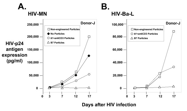
Results: We report here the use of non-infectious surface-engineered virion particles to modulate immunity against three infectious disease agents--human immunodeficiency virus type 1 (HIV-1), herpes simplex virus (HSV), and Influenza. Surface-engineering of particles are accomplished by genetic modification of the host cell surface that produces the egress budding viral particle. Human peripheral blood lymphocytes from healthy donors exposed to CD80/B7.1, CD86/B7.2, and/or antiCD3 single-chain antibody surface-engineered non-infectious HIV-1 and HSV-2 particles stimulate T cell proliferation, whereas particles released from non-modified host cells have no T cell stimulatory activity. In addition to T cell proliferation, HIV-based particles specifically suppress HIV-1 replication (both monocytotropic and lymphocytotropic strains) 55 to 96% and HSV-based particles specifically induce cross-reactive HSV-1/HSV-2 anti-herpes virus antibody production. Similar surface engineering of influenza-based particles did not modify the intrinsic ability of influenza particles to stimulate T cell proliferation, but did bestow on the engineered particles the ability to induce cross-strain anti-influenza antibody production.

Conclusion: We propose that non-infectious viral particles can be surface-engineered to produce antigen-presenting particles that mimic antigen-presenting cells to induce immune responses in human peripheral blood lymphocytes. The viral particles behave as "biological carriers" for recombinant proteins, thereby establishing a new therapeutic paradigm for molecular medicine.


Figures

Figure 1
Figure 1
Schematic representation of the retroviral vector constructions used to surface-modify particle-producing host cell lines. The detail construction of the vectors used in this report, pJDMT#6 (CD80/B7.1), pJDMT#19 (CD86/B7.2), and pJDMT#50 (antiCD3-sFv) are described in the Materials and Methods section.
Figure 2
Figure 2
Comparison of proliferation index (PI) in three donors (A, B, and C) human PBLs cultured with either PHA or particles surface-engineered with CD80, CD86, and antiCD3-sFv (B7+antiCD3). In Panel A, surface-engineered HSV-based particles are derived from HSV-2 infected genetically surface-modified Lof(11-10) cells (horizontal hatched bars). In Panel B, surface-engineered HIV-based particles are derived from genetically surface-modified 1119 cells that are chronically-expressing human immunodeficiency virus type-1 (gray-filled bars). The time course shown is 4, 6, 8, and 12 days for PHA-treated cultures; 4, 6, 8, 12, and 18 days for surface-engineered particle treated cultures. Proliferation Index establishes a proliferation ratio between exposed cultures and non-exposed cultures. PHA treated cultures are not exposed to particles. For PHA (black-filled bars), the proliferation value in the presence of PHA (i.e. Donor-A, 6 hour timepoint = 10,900 relative fluorescent units) is divided by untreated cultures not exposed to PHA (i.e. Donor-A, 6 hour timepoint = 2,500 relative fluorescent units); for B7+antiCD3, the proliferation value in the presence of B7+antiCD3 surface-engineered particles (i.e. Donor-A, 6 hour timepoint = 26,700 relative fluorescent units for HSV-2 in panel A; 11,000 relative fluorescent units for HIV-1 in panel B) is divided by the proliferation value observed with non-engineered viral-based particles (i.e. Donor-A, 6 hour timepoint = 2,300 relative fluorescent units for HSV-2 in panel A; 2,300 relative fluorescent units for HIV-1 in panel B). The remaining PI values are calculated in a similar fashion. Almost identical "background" values are observed for non-PHA exposed and non-engineered particles in Donors-A, -B, and -C cultures. Actual induced values can be calculated by multiplying the PI value by the "background" value. Particle preparations used in this figure were PEG-concentrated (200× for HIV; 25× for HSV) and inactivated to render them non-infectious.
Figure 3
Figure 3
Proliferation Index (PI) time course comparison in two donor (D and E) PBLs. Panel A: Non-engineered particles. For PHA (black-filled bars), the proliferation value in the presence of PHA (i.e. Donor-D, 6 hour timepoint = 4,000 relative fluorescent units) is divided by the value observed in untreated cultures (i.e. Donor-D, 6 hour timepoint = 2,000 relative fluorescent units); for HIV-1 (gray-filled bars), the proliferation value in the presence of non-engineered HIV-1 particles (i.e. Donor-D, 6 hour timepoint = 2,200 relative fluorescent units) is divided by the value observed in untreated cultures (i.e. Donor-D, 6 hour timepoint = 2,000 relative fluorescent units); for HSV-2 (horizontal hatched bars), the proliferation value in the presence of non-engineered HSV-2 particles (i.e. Donor-D, 6 hour timepoint = 2,000 relative fluorescent units) is divided by the value observed in untreated cultures (i.e. Donor-D, 6 hour timepoint = 2,000 relative fluorescent units); for Influenza A (PR8) (right-diagonal hatched bars), the proliferation value in the presence of non-engineered influenza-A particles (i.e. Donor-D, 6 hour timepoint = 26,000 relative fluorescent units) is divided by the value observed in untreated cultures (i.e. Donor-D, 6 hour timepoint = 2,000 relative fluorescent units); and for Influenza B (Russian) (left-diagonal hatched bars), the proliferation value in the presence of non-engineered influenza-B particles (i.e. Donor-D, 6 hour timepoint = 32,000 relative fluorescent units) is divided by the value observed in untreated cultures (i.e. Donor-D, 6 hour timepoint = 2,000 relative fluorescent units). Panel B: Surface-engineered influenza particles. For B7+antiCD3 surface-engineered influenza A (PR8) (right-diagonal hatched bars), the proliferation value in the presence of surface-engineered particles (i.e. Donor-D, 6 hour timepoint = 29,000 relative fluorescent units) is divided by the proliferation value observed with non-engineered influenza A particles (i.e. Donor-D, 6 hour timepoint = 26,000 relative fluorescent units); and for Influenza B (Russian) (left-diagonal hatched bars), the proliferation value in the presence of surface-engineered particles (i.e. Donor-D, 6 hour timepoint = 28,800 relative fluorescent units) is divided by the proliferation value observed with non-engineered influenza B particles (i.e. Donor-D, 6 hour timepoint = 32,000 relative fluorescent units). The time course shown in panels A and B for Donor-D is 4, 6, 10, 13, and 20 days; for Donor-E is 4, 10, 13, and 20 days. The remaining PI values are calculated in a similar fashion. Actual induced values can be calculated by multiplying the PI value by the "background" value. Particle preparations used in this figure were PEG-concentrated (200× for HIV; 25× for HSV; 40× for Influenza A/B) and inactivated to render them non-infectious.
Figure 4
Figure 4
Panel A: Proliferation Index (PI) time course comparison in three donors' (A, B, and C) PBLs cultured with either PHA or B7 surface-engineered HIV-1 based particles. For PHA (black-filled bars), the proliferation value in the presence of PHA (i.e. Donor-A, 6 hour timepoint = 10,900 relative fluorescent units) is divided by untreated cultures not exposed to PHA (i.e. Donor-A, 6 hour timepoint = 2,500 relative fluorescent units); for B7 (gray-filled bars) the proliferation value in the presence of B7 surface-engineered particles (i.e. Donor-A, 6 hour timepoint = 32,500 relative fluorescent units) is divided by the proliferation value observed with non-engineered HIV-based particles (i.e. Donor-A, 6 hour timepoint = 2,500 relative fluorescent). The time course shown is 4, 6, 8, 12, and 18 days for B7; 4, 6, 8, and 12 days for PHA. Particle preparations used in this panel were PEG-concentrated (200× for HIV; 25× for HSV) and inactivated to render them non-infectious. Panel B: Proliferation Index (PI) time course comparison in Donor-F PBLs cultured with HSV-2 based surface-engineered particles. For PHA (black-filled bars), the proliferation value in the presence of PHA (i.e. 8 hour timepoint = 9,000 relative fluorescent units) is divided by cultures not exposed to PHA (i.e. 8 hour timepoint = 2,600 relative fluorescent units); for AntiCD3 surface-engineered particles (tightly packed horizontal hatched gray bars), the proliferation value in the presence of AntiCD3 (i.e. 8 hour timepoint = 15,000 relative fluorescent units) is divided by the proliferation value observed with non-engineered HSV-based particles (i.e. 8 hour timepoint = 2,300 relative fluorescent units); for B7 surface-engineered particles (horizontal hatched gray bars), the proliferation value in the presence of B7 (i.e. 8 hour timepoint = 29,000 relative fluorescent units) is divided by the proliferation value observed with non-engineered HSV-based particles (i.e. 8 hour timepoint = 2,300 relative fluorescent units); for B7+antiCD3 surface-engineered particles (horizontal hatched bars), the proliferation value in the presence of B7+antiCD3 (i.e. 8 hour timepoint = 28,000 relative fluorescent units) is divided by the proliferation value observed with non-engineered HSV-based particles (i.e. 8 hour timepoint = 2,300 relative fluorescent units). Particle preparations used in this panel were from conditioned media and inactivated to render them non-infectious. Panel C: Proliferation Index (PI) time course comparison in Donor-F PBLs cultured with HSV-2 based surface-engineered particles. For PHA (black-filled bars), the proliferation value in the presence of PHA (i.e. 8 hour timepoint = 4,200 relative fluorescent units) is divided by cultures not exposed to PHA (i.e. 8 hour timepoint = 1,200 relative fluorescent units); for Heat-Inactivated B7+antiCD3 surface-engineered particles (tightly packed horizontal hatched gray lines), the proliferation value in the presence of heat-inactivated surface-engineered particles (i.e. 8 hour timepoint = 7,300 relative fluorescent units) is divided by the proliferation value observed with heat-inactivated non-engineered HSV-based particles (i.e. 8 hour timepoint = 6,500 relative fluorescent units); for Conditioned Media B7+antiCD3 (horizontal hatched bars), the proliferation value in the presence of conditioned media from surface-engineered particles (i.e. 8 hour timepoint = 30,000 relative fluorescent units) is divided by the proliferation value observed in conditioned media from non-engineered HSV-based particles (i.e. 8 hour timepoint = 2,700 relative fluorescent units); for Lyophilized room temperature stored B7+antiCD3 (checker bars), the proliferation value in the presence of the lyophilized surface-engineered particles (i.e. 8 hour timepoint = 28,000 relative fluorescent units) is divided by the proliferation value observed with lyophilized non-engineered HSV-based particles (i.e. 8 hour timepoint = 2,300 relative fluorescent units). The above data was obtained using Donor-F PBLs. PEG-concentrated B7+antiCD3 (brick bars) proliferation value was compared to Conditioned Media B7+antiCD3 (horizontal hatched bars) in Donor-H PBLs. The remaining PI values are calculated in a similar fashion. Almost identical "background" values are observed for non-PHA exposed and non-engineered particles in Donors-A, -B, -C, -F, and -H cultures. Actual induced values can be calculated by multiplying the PI value by the "background" value. Particle preparations used in this panel unless otherwise identified were from conditioned media and inactivated to render them non-infectious.
Figure 5
Figure 5
Surface-engineered HIV-based particle dependent inhibition of HIV-1 replication. Panel A: Lymphocytotropic HIV-1 MN p24 antigen expression in PHA-stimulated PBLs. Panel B: Monocytotropic HIV-1 Ba-L p24 antigen expression in PHA-stimulated PBLs. Donor-J cells were PHA-treated and exposed to either no particles (filled diamonds), non-engineered HIV-based particles (open squares), B7+antiCD3 surface-engineered HIV-based particles (open circles), or B7 surface-engineered HIV-based particles (open triangles). At day 3, cultures are washed and infectious HIV-1 is added – HIV-MN in Panel A and HIV-Ba-L in Panel B. Aliquots are removed at 3, 7, 12, and 17 days and HIV-1 encoded p24 antigen expression is determined by ELISA. Particle preparations used in this figure were PEG-concentrated and inactivated to render them non-infectious.

Similar articles

Cited by

References

    1. Dorrell Lucy. Therapeutic immunization strategies for the control of HIV-1. Vaccine. 2005;4:513–520. - PubMed
    1. Nolan D, Reiss P, Mallal S. Adverse effects of antiretroviral therapy for HIV infection: a review of selected topics. Drug Saf. 2005;4:201–218. - PubMed
    1. Penzak SR, Formentini E, Alfaro RM, Long M, Natarajan V, Kovacs J. Prednisolone Pharmacokinetics in the Presence and Absence of Ritonavir After Oral Prednisone Administration to Healthy Volunteers. JAIDS. 2005;40:573–580. - PubMed
    1. Rosenberg SA, Dudley ME. Cancer regression in patients with metastatic melanoma after the transfer of autologous antitumor lymphocytes. Proc Natl Acad Sci USA. 2004;101:14639–14645. doi: 10.1073/pnas.0405730101. - DOI - PMC - PubMed
    1. Dermime S, Armstrong A, Hawkins RE, Stern PL. Cancer vaccines and immunotherapy. Brit Med. 2002;62:149–162. doi: 10.1093/bmb/62.1.149. - DOI - PubMed

MeSH terms