. 2007 May 15;21(10):1217-30.
doi: 10.1101/gad.1528307.

Dissociation of eIF1 from the 40S ribosomal subunit is a key step in start codon selection in vivo

Affiliations

Dissociation of eIF1 from the 40S ribosomal subunit is a key step in start codon selection in vivo

Yuen-Nei Cheung et al. Genes Dev. .

Abstract

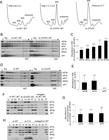
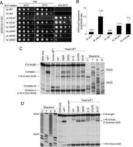
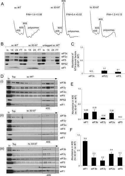
Selection of the AUG start codon is a key step in translation initiation requiring hydrolysis of GTP in the eIF2*GTP*Met-tRNA(i)(Met) ternary complex (TC) and subsequent P(i) release from eIF2*GDP*P(i). It is thought that eIF1 prevents recognition of non-AUGs by promoting scanning and blocking P(i) release at non-AUG codons. We show that Sui(-) mutations in Saccharomyces cerevisiae eIF1, which increase initiation at UUG codons, reduce interaction of eIF1 with 40S subunits in vitro and in vivo, and both defects are diminished in cells by overexpressing the mutant proteins. Remarkably, Sui(-) mutation ISQLG(93-97)ASQAA (abbreviated 93-97) accelerates eIF1 dissociation and P(i) release from reconstituted preinitiation complexes (PICs), whereas a hyperaccuracy mutation in eIF1A (that suppresses Sui(-) mutations) decreases the eIF1 off-rate. These findings demonstrate that eIF1 dissociation is a critical step in start codon selection, which is modulated by eIF1A. We also describe Gcd(-) mutations in eIF1 that impair TC loading on 40S subunits or destabilize the multifactor complex containing eIF1, eIF3, eIF5, and TC, showing that eIF1 promotes PIC assembly in vivo beyond its important functions in AUG selection.


Figures

Figure 1.
Figure 1.
Gcd mutations in eIF1 reduce TC loading on 40S subunits in vitro. (A) Space-filling model (left) or ribbons depiction (right) of human eIF1 (PDB file 2IF1) highlighting residues corresponding to mutations in yeast eIF1 with Gcd or Sui phenotypes. (B) Analysis of Gcd phenotypes in gcn2Δ strains. Serial dilutions of yeast cells were incubated for 2 d at 30°C on SC lacking uracil and leucine (SC-UL) (left) and 4 d on SC-UL lacking histidine and containing 20 mM or 5 mM 3-AT (right). (Rows 25) Strains harboring wild-type (JCY103) or 9,12 (JCY115) sc His-SUI1 alleles and either vector or hc TC plasmid p1780-IMT. (Rows 6,7) Strains with hc wild-type (JCY105) or G107R His-SUI1 alleles (JCY211) and sc SUI1+ plasmid p1200. (Rows 810) Strains with wildtype (JCY103), 93–97 (JCY137), or D83G (JCY221) His-SUI1 alleles on sc or hc plasmids. (Row 1) Isogenic GCN2 strain H1642. (C) Expression of GCN4-lacZ (with all four uORFs) in the gcn2Δ strains described in B grown in SC-L or SC-UL. β-Galactosidase activities (nanomoles of o-nitrophenyl-β-D-galactopyranoside cleaved per minute per microgram of protein) were measured in WCEs, and the mean and standard error (SE) from three or more measurements on six independent transformants are plotted. (D) GCN2 his4-303 strains with His-SUI1 alleles wild type (JCY145), 93–97 (JCY189), hc wild type (JCY149), or hc G107R (JCY197), and harboring the GCN4-lacZ plasmid (with all four ORFs, from plasmid p180) were grown in SC-UL also lacking Ile/Val (SC-ULIV) for 6 h with or without sulfometuron (0.5 μg/mL), and β-galactosidase activities were measured. (E) eIF1 mutations 9,12 and G107R reduce the rate of TC loading on reconstituted PICs. Mutant or wild-type eIF1 was mixed with preformed TC (eIF2•GDPNP•35S- Met-tRNAiMet), eIF1A, 40S ribosomes, and mRNA, and the fraction of labeled 35S-Met-tRNAiMet bound to 40S subunits was measured over time by native gel electrophoresis. eIF1 was saturating (1 μM) in all cases. (F) Mutation 93–97, but not 9,12 or G107R, significantly reduces eIF1 affinity for 48S PICs. TAMRA-labeled wild-type or mutant eIF1 was mixed with 40S subunits, eIF1A, TC, and mRNA, and the increase in fluorescence anisotropy at equilibrium (yielding the fraction of eIF1 bound to 43S•mRNA complexes) was measured at different concentrations of the complex.
Figure 2.
Figure 2.
Gcd eIF1 mutation G107R reduces translation initiation and disrupts MFC integrity. (A,B) Effects on translation initiation and PIC levels in vivo. Strains JCY105, JCY211, and JCY197 were grown in SC-UL or SC-L and cross-linked with 1% (v/v) HCHO for 1 h. (A) WCEs were resolved by sedimentation through 4.5%–45% sucrose gradients at 39,000 rpm for 2.5 h and scanned at A254 to determine polysome/monosome ratios (P/M, mean ± SE, n = 3; [*] p < 0.05). (B) Fractions from a 7.5%–30% gradient centrifuged at 41,000 rpm for 5 h and 1% of each WCE (In) were subjected to Western analysis. Fractions containing free 40S subunits are boxed. (C) Initiation factor binding to 40S subunits for the experiment in B and two replicates was quantified by calculating the ratio of eIF signals in 40S fractions of the mutant versus wild-type gradients for each factor. The results plotted are means ± SEs (n = 3). (D) The analysis described in B was performed using strains JCY149 and JCY197, and the 40S fractions were pooled, resolved on a second gradient, and subjected to Western analysis. (E) eIF binding to 40S subunits in the resedimentation experiment in D and two replicates were quantified, plotting the means ± SEs (n = 3). (F) eIF1 mutation G107R impairs MFC integrity in vivo. Nickel chelation chromatography of WCEs from strains JCY105, JCY211, and JCY255. Eluted proteins were subjected to Western analysis. (In) 3% of WCEs input; (1× and 2×) 15% and 30% of eluates; (FT) 1% of flowthrough. (G) For the experiment in F and two replicates, the amounts of eIFs in the eluates were quantified and normalized to the amounts of coeluting His6-eIF1, and the ratio of the normalized values for hc G107R/WT versus hc WT/WT strains is plotted (mean values and SEs; n = 3). (H) WCEs of strains JCY103, JCY115, and JCY253 were analyzed by nickel chelation chromatography as in F.
Figure 3.
Figure 3.
eIF1 mutations D83G, Q84P, and 93–97 reduce stringent AUG selection in vivo and in vitro. (A) GCN2 his4-303 strains containing His-SUI1 alleles 93–97, D83G, and Q84P were grown on SC-L for 2 d at 30°C or 37°C, and on SC-LH (−His) for 7 d to reveal Sui phenotypes. (B) Quantification of Sui phenotypes. Strains from A with HIS4-lacZ reporters with AUG (p367) or UUG (p391), respectively, were grown in SD with His and Trp at 30°C, and β-galactosidase activities were measured in WCEs. The mean ratios of UUG to AUG reporter expression are shown with SEs, from three experiments on six independent transformants. (C) Scanning defects of eIF1 mutants revealed by toeprinting analysis of 48S PICs assembled on native β-globin mRNA in reactions containing mammalian 40S ribosomes, eIF1A, eIF2, eIF3, eIF4A, eIF4B, eIF4F, GTP, and Met-tRNAiMet in the absence or presence of human eIF1 or various yeast wild-type or mutant eIF1 proteins. (D) Toeprinting analysis of 48S complexes formed on a (CAA)nGUS mRNA containing an AUG 1 nt downstream from the 5′ end, as in C.
Figure 4.
Figure 4.
Overexpression of eIF1 mutant 93–97 suppresses its growth defect and partially rescues PIC assembly in vivo. (A) eIF1 mutation 93–97 impairs translation in vivo in a manner suppressed by overexpression. Analysis of polysome profiles, conducted as in Figure 2A, on strains JCY145, JCY189, and JCY193. (B,C) Mutant 93–97 impairs MFC integrity. Nickel chelation chromatography of WCEs from strains JCY145, JCY189, and JCY261 conducted as in Figure 2F,G. (D) 93–97 affects native PIC assembly. Resedimentation analysis of native PICs quantified as described in Figure 2D. (E,F) Quantification of eIF binding to 40S subunits in the experiment shown in D and two replicate experiments, as in Figure 2E.
Figure 5.
Figure 5.
Sui mutation 93–97 increases the rates of eIF1 dissociation and Pi release from eIF2•GDP•Pi in reconstituted 48S PICs. (A) Reduced affinity of the 93–97 mutant for 43S•UUG complexes demonstrated in vitro as described in Table 2. (B) 93–97 accelerates dissociation of eIF1 from 43S•mRNA complexes. 43S complexes reconstituted with TC, eIF1A-fl, and mutant or wild-type eIF1-TAMRA were mixed rapidly with mRNA (AUG) and excess unlabeled eIF1, and the increase in eIF1A-fl fluorescence (caused by decrease in FRET efficiency) was monitored by stopped-flow fluorometry. (C,D) 93–97 accelerates the Pi-release phase of GTP hydrolysis. 43S complexes (with [γ-32P]GTP in the TC) were mixed with eIF5, excess unlabeled GDP, and model mRNA containing AUG (C) or UUG (D) start codons in a rapid quench apparatus, and reactions were quenched at the indicated times with EDTA. The extent of GTP hydrolysis was measured by separating free 32Pi from [γ-32P]GTP by gel electrophoresis followed by PhosphorImager analysis. (E) FL-17–21 mutation in eIF1A decreases the rate of eIF1 dissociation from 43S•mRNA complexes. Kinetic experiments conducted as in B using mutant or wild-type forms of eIF1A-fl and wild-type eIF1-TAMRA in 43S•mRNA (AUG) complexes. For wild-type eIF1A, the rate constants (k) and amplitudes (amp) of the fast (conformational change) and slow (eIF1 release) phases, respectively, are k1 = 9.2 ± 0.3 sec−1, amp1 = 0.16 ± 0.002 and k2 = 0.32 ± 0.003 sec−1, amp2 = 0.83 ± 0.003. The corresponding values for FL-17–21 mutant are k1 = 1.0 ± 0.4 sec−1, amp1 = 0.19 ± 0.03 and k2 = 0.15 ± 0.007 sec−1, amp2 = 0.81 ± 0.09.

Comment in

Similar articles

Cited by

References

    1. Algire M.A., Maag D., Savio P., Acker M.G., Tarun S.Z., Jr., Sachs A.B., Asano K., Nielsen K.H., Olsen D.S., Phan L., Maag D., Savio P., Acker M.G., Tarun S.Z., Jr., Sachs A.B., Asano K., Nielsen K.H., Olsen D.S., Phan L., Savio P., Acker M.G., Tarun S.Z., Jr., Sachs A.B., Asano K., Nielsen K.H., Olsen D.S., Phan L., Acker M.G., Tarun S.Z., Jr., Sachs A.B., Asano K., Nielsen K.H., Olsen D.S., Phan L., Tarun S.Z., Jr., Sachs A.B., Asano K., Nielsen K.H., Olsen D.S., Phan L., Sachs A.B., Asano K., Nielsen K.H., Olsen D.S., Phan L., Asano K., Nielsen K.H., Olsen D.S., Phan L., Nielsen K.H., Olsen D.S., Phan L., Olsen D.S., Phan L., Phan L., et al. Development and characterization of a reconstituted yeast translation initiation system. RNA. 2002;8:382–397. - PMC - PubMed
    1. Algire M.A., Maag D., Lorsch J.R., Maag D., Lorsch J.R., Lorsch J.R. π release from eIF2, not GTP hydrolysis, is the step controlled by start-site selection during eukaryotic translation initiation. Mol. Cell. 2005;20:251–262. - PubMed
    1. Asano K., Phan L., Anderson J., Hinnebusch A.G., Phan L., Anderson J., Hinnebusch A.G., Anderson J., Hinnebusch A.G., Hinnebusch A.G. Complex formation by all five homologues of mammalian translation initiation factor 3 subunits from yeast Saccharomyces cerevisiae. J. Biol. Chem. 1998;273:18573–18585. - PubMed
    1. Asano K., Clayton J., Shalev A., Hinnebusch A.G., Clayton J., Shalev A., Hinnebusch A.G., Shalev A., Hinnebusch A.G., Hinnebusch A.G. A multifactor complex of eukaryotic initiation factors eIF1, eIF2, eIF3, eIF5, and initiator tRNAMet is an important translation initiation intermediate in vivo. Genes & Dev. 2000;14:2534–2546. - PMC - PubMed
    1. Asano K., Shalev A., Phan L., Nielsen K., Clayton J., Valášek L., Donahue T.F., Hinnebusch A.G., Shalev A., Phan L., Nielsen K., Clayton J., Valášek L., Donahue T.F., Hinnebusch A.G., Phan L., Nielsen K., Clayton J., Valášek L., Donahue T.F., Hinnebusch A.G., Nielsen K., Clayton J., Valášek L., Donahue T.F., Hinnebusch A.G., Clayton J., Valášek L., Donahue T.F., Hinnebusch A.G., Valášek L., Donahue T.F., Hinnebusch A.G., Donahue T.F., Hinnebusch A.G., Hinnebusch A.G. Multiple roles for the carboxyl terminal domain of eIF5 in translation initiation complex assembly and GTPase activation. EMBO J. 2001;20:2326–2337. - PMC - PubMed

Publication types

MeSH terms

Substances