Comparative Study
. 2007 Aug;21(8):1861-76.
doi: 10.1210/me.2006-0101. Epub 2007 May 15.

The HER4 cytoplasmic domain, but not its C terminus, inhibits mammary cell proliferation

Affiliations
Comparative Study

The HER4 cytoplasmic domain, but not its C terminus, inhibits mammary cell proliferation

Shu-Mang Feng et al. Mol Endocrinol. 2007 Aug.

Abstract

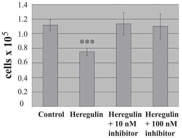
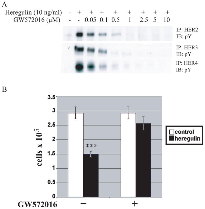
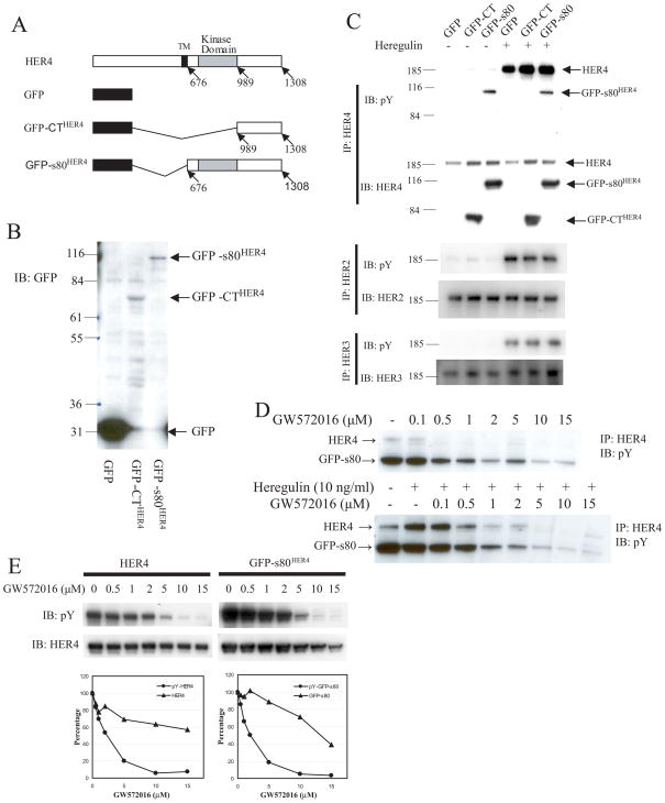
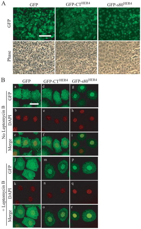
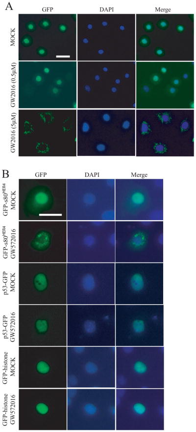
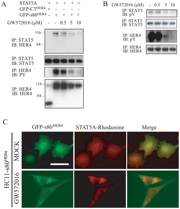
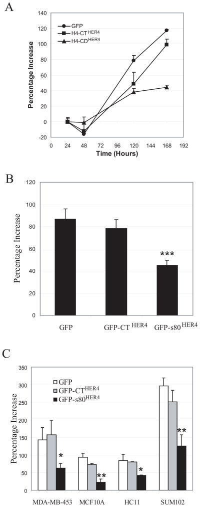
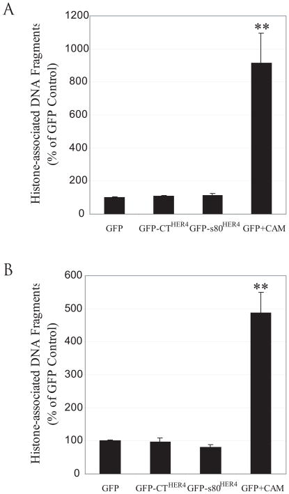
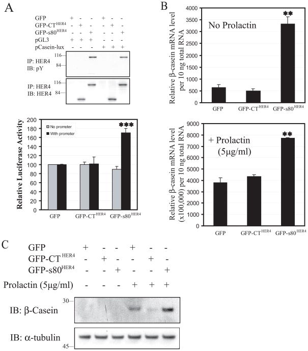
Unlike the proliferative action of other epidermal growth factor (EGF) receptor family members, HER4/ErbB4 is often associated with growth-inhibitory and differentiation signaling. These actions may involve HER4 two-step proteolytic processing by intramembraneous gamma-secretase, releasing the soluble, intracellular 80-kDa HER4 cytoplasmic domain, s80HER4. We demonstrate that pharmacological inhibition of either gamma-secretase activity or HER4 tyrosine kinase activity blocked heregulin-dependent growth inhibition of SUM44 breast cancer cells. We next generated breast cell lines stably expressing GFP-s80HER4 [green fluorescent protein (GFP) fused to the N terminus of the HER4 cytoplasmic domain, residues 676-1308], GFP-CT(HER4) (GFP fused to N terminus of the HER4 C-terminus distal to the tyrosine kinase domain, residues 989-1308), or GFP alone. Both GFP-s80HER4 and GFP-CTHER4 were found in the nucleus, but GFP-s80HER4 accumulated to a greater extent and sustained its nuclear localization. s80HER4 was constitutively tyrosine phosphorylated, and treatment of cells with a specific HER family tyrosine kinase inhibitor 1) blocked tyrosine phosphorylation; 2) markedly diminished GFP-s80HER4 nuclear localization; and 3) reduced signal transducer and activator of transcription (STAT)5A tyrosine phosphorylation and nuclear localization as well as GFP-s80HER4:STAT5A interaction. Multiple normal mammary and breast cancer cell lines, stably expressing GFP-s80HER4 (SUM44, MDA-MB-453, MCF10A, SUM102, and HC11) were growth inhibited compared with the same cell line expressing GFP-CTHER4 or GFP alone. The s80HER4-induced cell number reduction was due to slower growth because rates of apoptosis were equivalent in GFP-, GFP-CTHER4-, and GFP-s80HER4-expressing cells. Lastly, GFP-s80HER4 enhanced differentiation signaling as indicated by increased basal and prolactin-dependent beta-casein expression. These results indicate that surface HER4 tyrosine phosphorylation and ligand-dependent release of s80HER4 are necessary, and s80HER4 signaling is sufficient for HER4-dependent growth inhibition.


Figures

Fig. 1
Fig. 1. Inhibition of γ-secretase blocks heregulin-induced growth inhibition
SUM44 cells grown in fully complemented medium were treated with heregulin (10 ng/ml) or not in the presence or absence of HEDI at the concentrations shown for 6 days, then counted. Mean ± SD of triplicates are shown, representative of 3 independent experiments. Student’s t-test was used for statistical analysis. ***, P < 0.001 versus control.
Fig. 2
Fig. 2. Inhibition of HER4 tyrosine phosphorylation blocks heregulin-induced growth inhibition
A, SUM44 cells were cultured for 6 days, treated with different doses of GW572016 for 1 h, and then with or without heregulin (10 ng/ml) for 15 minutes. The cells were lysed and the lysates were immunoprecipated with anti-HER2, anti-HER3, or anti-HER4 antibody and blotted with anti-phosphotyrosine antibody. B, SUM44 cells were grown with or without heregulin (10 ng/ml) in the presence or absence of GW572016 (1 μM) for 6 days, at which time the number of cells were counted. Mean ± SD of triplicates are shown, representative of 3 experiments. ***, P < 0.001 versus control.
Fig. 3
Fig. 3. Ectopic expression of GFP-s80HER4 and GFP-CTHER4
A, Schematic representation of full-length HER4, GFP, GFP-CTHER4, and GFP-s80 HER4. B, Western analysis using anti-GFP antibody of lysates from SUM44 cells infected with retrovirus encoding GFP, GFP-CTHER4, or GFP-s80HER4. C, SUM44 cells stably expressing GFP, GFP-CTHER4, or GFP-s80HER4 were treated with or without heregulin (10 ng/ml) for 15 minutes, and lysed. The lysates were immunoprecipated with anti-HER2, anti-HER3, or anti-HER4 antibody and blotted with anti-HER2, anti-HER3, or anti-HER4 antibody respectively, or with anti-phosphotyrosine antibody. D, HC11 cells stably expressing GFP, GFP-CTHER4, or GFP-s80HER4 were treated with ± increasing concentrations of GW572016 for 1 h (top panel), or treated with different concentration of GW572016 for 1 h, then ± heregulin (10 ng/ml) for 15 minutes (bottom panel), and lysed. The lysates were immunoprecipated with anti-HER4 antibody and blotted with anti-phosphotyrosine antibody. E, COS-7 cells were transfected with full-length HER4 or GFP-s80HER4. Twenty-four h after transfection, the cells were treated with ± increasing concentrations of GW572016 for 1 h, lysed, and analysed for the level of tyrosine phosphorylation of HER4 or GFP-s80HER4 by using anti-phosphotyrosine antibody, and the level of HER4 or GFP-s80HER4 by using anti-HER4 antibody. Densitometry is shown in arbitrary units.
Fig. 4
Fig. 4. Localization of GFP, GFP-CTHER4, and GFP-s80HER4 in HC11 cells
A, Live cell microscopy showing the localization of GFP fluorescence in HC11 cells stably expressing GFP, GFP-CTHER4, or GFP-s80HER4. Phase contrast is shown in lower panels. Bar, 100 μm, applies to each image. B, HC11 cells stably expressing GFP (a-c, j-l), GFP-CTHER4 (d-f, m-o) or GFP-s80HER4 (g-i, p-r), were treated without (a-i), or with (j-r) 20 ng/ml of leptomycin B for 24 h, then analyzed for GFP fluorescence by confocal microscopy. Subcellular distribution of GFP (a, j), GFP-CT (d, m), GFP-s80 (g, p), and DAPI (b, e, h, k, n, q) were visualized and captured. The merged pictures were also shown (c, f, i, l, o, r). Bar, 30 μm, applies to each image.
Fig. 5
Fig. 5. Effect of GW572016 on the localization of GFP-s80HER4 in HC11 cells
A, HC11 cells stably expressing GFP-s80HER4 were treated without, or with 0.5 μM or 5 μM GW572016 for 24 h. Cells on the cover slip were fixed, stained with DAPI, then analyzed for GFP and DAPI fluorescence. The merged pictures were also shown. Bar, 30 μm, applies to each image. B, COS-7 cells were transfected with GFP-s80HER4, p53-GFP, or GFP-histone, and treated without or with 10 μM GW572016 for 24 h. Then, the cells were fixed, stained with DAPI, and analyzed for GFP and DAPI fluorescence. The merged pictures were also shown. Bar, 30 μm, applies to each image.
Fig. 6
Fig. 6. Effect of GW572016 on STAT5A and GFP-s80HER4 co-immunoprecipitation, STAT5A tyrosine phosphorylation and STAT5A nuclear localization
A, COS-7 cells were co-transfected with STAT5A and GFP-CTHER4 or STAT5A and GFP-s80HER4, and treated without or with increasing concentrations of GW572016 for 40 hours. Cells were lysed with regular lysis buffer (137mM NaCl), and lysates immunoprecipitated with anti-HER4 or anti-STAT5 antibody. Immunoprecipitates were subject to gel electrophoresis and transferred and blotted with anti-HER4, anti-STAT5, or anti-phosphotyrosine antibody as indicated. B, COS-7 cells were co-transfected with GFP-s80HER4 and STAT5A, and treated without or with different concentrations of GW572016 for 40 hours. Cells were lysed in high salt (500 mM NaCl) lysis buffer, and the lysates were immunoprecipitated with anti-HER4 or anti-STAT5 antibody, electrophoresed, transferred, and blotted with anti-phosphotyrosine, anti-STAT5 or anti-HER4 antibody. C, HC11 cells stably expressing GFP-s80HER4 were treated without or with GW572016 (5 μM) for 24 h. Cells were fixed and visualized for GFP or rhodamine (using a rhodamine-labeled second antibody following the first antibody-antiSTAT5). The merged pictures are also shown. Bar, 30 μm, applies to each image.
Fig. 7
Fig. 7. Ectopic expression of GFP-s80HER4, but not GFP-CTHER4, decreases cell proliferation
A, Time course of cell proliferation of SUM44 cells stably expressing GFP, GFP-CTHER4, or GFP-s80HER4. SUM44 cells (5000 cells per well) stably expressing GFP, GFP-CTHER4, or GFP-s80HER4 were plated in 96-well plates and cultured. At indicated time points, the relative cell numbers (OD values) were measured using MTS assay. Percentage increased OD value at each time point over the 20h time point are shown (Mean ± S.E.M. of triplicates). B, Cell proliferation of SUM44 cells stably expressing GFP, GFP-CTHER4, or GFP-s80HER4 after 7 days incubation. The percentages of increased OD value at the end of the incubation (7 days) to that at 20 hours are shown. The error bar represented S.E.M. of five independent experiments, each analyzed in triplicate. Student’s t-test was used for statistical analysis. ***, P < 0.001 versus GFP control. C, MTS assays showing cell proliferation of MDA-MB-453, MCF10, HC11 and SUM 102 cells stably expressing GFP, GFP-CTHER4 or GFP-s80HER4. OD values of the cells stably expressing GFP, GFP-CTHER4 or GFP-s80HER4 were determined at 20h and the end of the cell culture (MDA-MB-453, 5 days; MCF10A, 8 days; HC11, 3 days; SUM102, 4 days) using MTS assay. The percentages of increased OD value at the end of the incubation to that at 20 h are shown (the error bar represented S.E.M of triplicate samples). *, P < 0.05; **, P < 0.01 versus GFP control.
Fig. 8
Fig. 8. Stable expression of GFP-s80HER4 does not induce apoptosis
A, SUM44 cells stably expressing GFP, GFP-CTHER4 or GFP-S80HER4 were cultured in serum-free medium for 7 days. GFP expressing cells were also treated with 1 μM of camptothecin for 24 h before harvesting the cells as a positive control. Equal cell numbers were analyzed for apoptosis by ELIZA, which quantitatively measures cytoplasmic histone-associated DNA fragments. Results are expressed as mean±S.E.M. of three independent experiments; each sample being analyzed in duplicate. The Student’s t-test was used for statistical analysis. **, P = 0.006 versus GFP control. B, HC-11 cells stably expressing GFP, GFP-CTHER4 or GFP-S80HER4 were analyzed for apoptosis after 3 days as described above. A second plate of GFP expressing cells was treated with 1 μM of camptothecin for the final 24 h of the incubation. **, P = 0.04 versus GFP control.
Fig. 9
Fig. 9. GFP-s80HER4, but not GFP-CTHER4, activates β–casein promoter and increases β–casein mRNA and protein expression
A, HC11 cells transfected with plasmids containing GFP, GFP-CTHER4 or GFP-S80HER4, and pβcasein-lux, a reporter construct in which a human β–casein promoter was fused to the upstream of luciferase reporter gene, or pGL3 vector without β–casein promoter, were lysed 48 h after transfection. The lysates were immunoprecipitated with anti-HER4 antibody and blotted with anti-phosphotyrosine antibody, and anti-HER4 antibody (top panel). The lysates from above were also used to determine luciferase activity by luciferase assay system (Promega). Results are expressed as mean±S.E.M. of three independent experiments with each sample being analyzed in duplicate (bottom panel). The Student’s t-test was used for statistical analysis. ***, P < 0.001 versus GFP control or GFP-CTHER4. B, β–casein mRNA levels as determined by quantitative reverse transcription-PCR (qRT-PCR). HC11 cells stably expressing GFP, GFP-CTHER4, or GFP-s80HER4 were cultured in normal culture medium for 2 days, changed to serum-free complemented medium containing 5 μg/ml insulin for 2 days, then treated with or without 5 μg/ml prolactin in the complemented medium for 2 days. Total RNA was extracted and qRT-PCR was performed using β–casein specific fluorescence-labeled oligonucleotide probes (the error bar represented S.E.M of triplicate samples). **, P ≤ 0.01 versus GFP control. C, HC11 cells stably expressing GFP, GFP-CTHER4, or GFP-s80HER4 were cultured as above, and treated with or without 5 μg/ml prolactin in the complemented medium for 2 days. The cells were lysed and the lysates were immuno-blotted with anti-β-casein or anti-α-tubulin antibody.

Similar articles

Cited by

References

    1. Lacenere CJ, Sternberg PW. Regulation of EGF receptor signaling in the fruitfly D. melanogaster and the nematode C. elegans. Breast Dis. 2000;11:19–30. - PubMed
    1. Yarden Y, Sliwkowski MX. Untangling the ErbB signalling network. Nat Rev Mol Cell Biol. 2001;2:127–137. - PubMed
    1. Earp HS, 3rd, Calvo BF, Sartor CI. The EGF receptor family--multiple roles in proliferation, differentiation, and neoplasia with an emphasis on HER4. Trans Am Clin Climatol Assoc. 2003;114:315–333. - PMC - PubMed
    1. Roskoski R., Jr The ErbB/HER receptor protein-tyrosine kinases and cancer. Biochem Biophys Res Commun. 2004;319:1–11. - PubMed
    1. Holbro T, Hynes NE. ErbB receptors: directing key signaling networks throughout life. Annu Rev Pharmacol Toxicol. 2004;44:195–217. - PubMed

Publication types

MeSH terms