

Abnormal melatonin synthesis in autism spectrum disorders
- PMID: 17505466
- PMCID: PMC2199264
- DOI: 10.1038/sj.mp.4002016
Abnormal melatonin synthesis in autism spectrum disorders
Abstract
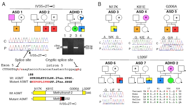
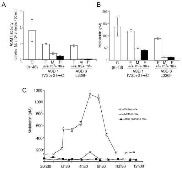
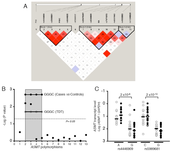
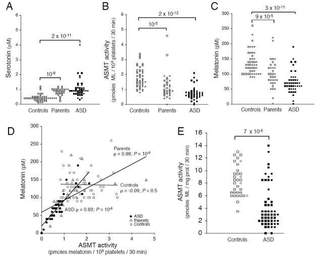
Melatonin is produced in the dark by the pineal gland and is a key regulator of circadian and seasonal rhythms. A low melatonin level has been reported in individuals with autism spectrum disorders (ASD), but the underlying cause of this deficit was unknown. The ASMT gene, encoding the last enzyme of melatonin synthesis, is located on the pseudo-autosomal region 1 of the sex chromosomes, deleted in several individuals with ASD. In this study, we sequenced all ASMT exons and promoters in individuals with ASD (n=250) and compared the allelic frequencies with controls (n=255). Non-conservative variations of ASMT were identified, including a splicing mutation present in two families with ASD, but not in controls. Two polymorphisms located in the promoter (rs4446909 and rs5989681) were more frequent in ASD compared to controls (P=0.0006) and were associated with a dramatic decrease in ASMT transcripts in blood cell lines (P=2 x 10(-10)). Biochemical analyses performed on blood platelets and/or cultured cells revealed a highly significant decrease in ASMT activity (P=2 x 10(-12)) and melatonin level (P=3 x 10(-11)) in individuals with ASD. These results indicate that a low melatonin level, caused by a primary deficit in ASMT activity, is a risk factor for ASD. They also support ASMT as a susceptibility gene for ASD and highlight the crucial role of melatonin in human cognition and behavior.
Figures
Similar articles
-
Sequencing ASMT identifies rare mutations in Chinese Han patients with autism.PLoS One. 2013;8(1):e53727. doi: 10.1371/journal.pone.0053727. Epub 2013 Jan 17. PLoS One. 2013. PMID: 23349736 Free PMC article.
-
Genetic and functional abnormalities of the melatonin biosynthesis pathway in patients with bipolar disorder.Hum Mol Genet. 2012 Sep 15;21(18):4030-7. doi: 10.1093/hmg/dds227. Epub 2012 Jun 13. Hum Mol Genet. 2012. PMID: 22694957
-
Single-nucleotide polymorphisms and mRNA expression for melatonin synthesis rate-limiting enzyme in recurrent depressive disorder.J Pineal Res. 2010 May;48(4):311-7. doi: 10.1111/j.1600-079X.2010.00754.x. J Pineal Res. 2010. PMID: 20433639
-
[Association of polymorphic variants of DDC (AADC), AANAT and ASMT genes encoding enzymes for melatonin synthesis with the higher risk of neuropsychiatric disorders].Zh Nevrol Psikhiatr Im S S Korsakova. 2021;121(5):151-157. doi: 10.17116/jnevro2021121041151. Zh Nevrol Psikhiatr Im S S Korsakova. 2021. PMID: 34184492 Review. Russian.
-
Autism spectrum disorder (ASD): Disturbance of the melatonin system and its implications.Biomed Pharmacother. 2020 Oct;130:110496. doi: 10.1016/j.biopha.2020.110496. Epub 2020 Jul 15. Biomed Pharmacother. 2020. PMID: 32682113 Review.
Cited by
-
Is sleep essential for neural plasticity in humans, and how does it affect motor and cognitive recovery?Neural Plast. 2013;2013:103949. doi: 10.1155/2013/103949. Epub 2013 Jun 11. Neural Plast. 2013. PMID: 23840970 Free PMC article. Review.
-
Molecular Pathology and Pharmacological Treatment of Autism Spectrum Disorder-Like Phenotypes Using Rodent Models.Front Cell Neurosci. 2018 Nov 20;12:422. doi: 10.3389/fncel.2018.00422. eCollection 2018. Front Cell Neurosci. 2018. PMID: 30524240 Free PMC article. Review.
-
Parent-of-origin effects in autism identified through genome-wide linkage analysis of 16,000 SNPs.PLoS One. 2010 Sep 2;5(9):e12513. doi: 10.1371/journal.pone.0012513. PLoS One. 2010. PMID: 20824079 Free PMC article.
-
A systematic review of common genetic variation and biological pathways in autism spectrum disorder.BMC Neurosci. 2021 Oct 9;22(1):60. doi: 10.1186/s12868-021-00662-z. BMC Neurosci. 2021. PMID: 34627165 Free PMC article.
-
Sleep disturbances in autism spectrum disorder: Animal models, neural mechanisms, and therapeutics.Neurobiol Sleep Circadian Rhythms. 2023 Apr 26;14:100095. doi: 10.1016/j.nbscr.2023.100095. eCollection 2023 May. Neurobiol Sleep Circadian Rhythms. 2023. PMID: 37188242 Free PMC article. Review.
References
-
- Axelrod J. The pineal gland: a neurochemical transducer. Science. 1974;184:1341–1348. - PubMed
-
- Brzezinski A. Melatonin in humans. N Engl J Med. 1997;336:186–195. - PubMed
-
- Arendt J. Melatonin, circadian rhythms, and sleep. N Engl J Med. 2000;343:1114–1116. - PubMed
-
- Saper CB, Scammell TE, Lu J. Hypothalamic regulation of sleep and circadian rhythms. Nature. 2005;437:1257–1263. - PubMed
-
- Simonneaux V, Ribelayga C. Generation of the melatonin endocrine message in mammals: a review of the complex regulation of melatonin synthesis by norepinephrine, peptides, and other pineal transmitters. Pharmacol Rev. 2003;55:325–395. - PubMed
Publication types
MeSH terms
Substances
LinkOut - more resources
Full Text Sources
Other Literature Sources
Molecular Biology Databases
