

Application of vacuum-assisted therapy in postoperative ascitic fluid leaks: an integral part of multimodality wound management in cirrhotic patients
- PMID: 17505531
- PMCID: PMC1855689
Application of vacuum-assisted therapy in postoperative ascitic fluid leaks: an integral part of multimodality wound management in cirrhotic patients
Abstract
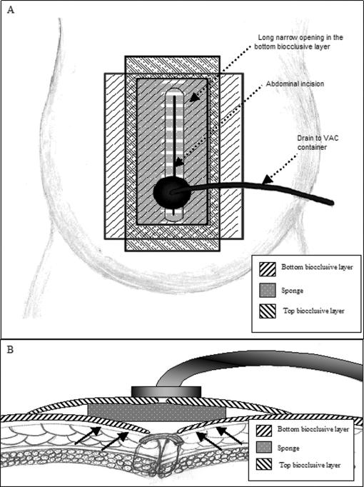
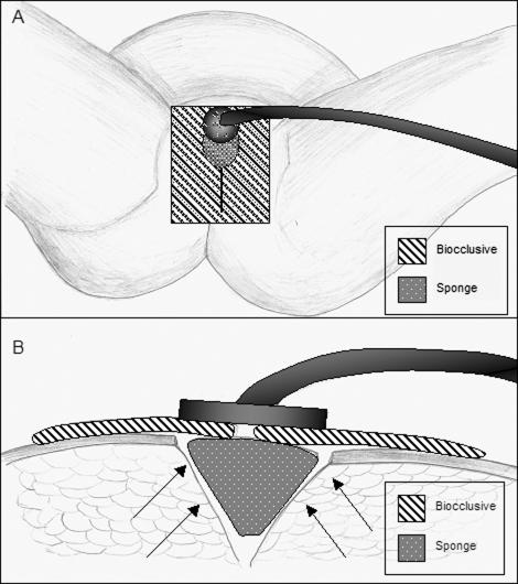
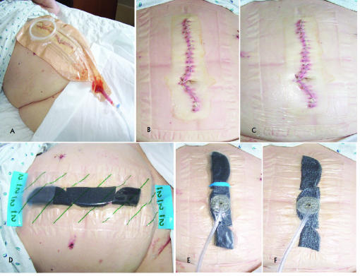
Surgery in patients with hepatic cirrhosis and ascites is associated with significant morbidity, including poor wound healing. Postoperative management of abdominal and perineal wounds in these patients poses a unique challenge owing to increased intra-abdominal pressure, risk for peritonitis, and ascitic fluid leakage. Vacuum-assisted closure (VAC) therapy reportedly improves angiogenesis and epithelialization, controls bacterial contamination, and removes excess tissue fluid. We present 4 cases of successful management of intractable postoperative ascitic fluid leaks utilizing VAC-based techniques. In one case, closure of a profusely draining perineal wound following an abdominoperineal resection was accomplished within 5 days of specialized VAC dressing application. In the other 3 cases, refractory drainage from midline laparotomy incision was successfully managed with the use of VAC therapy. In all 4 cases, the VAC-based system was effective in controlling drainage of ascites and subsequently sealing the wounds. Postoperative use of VAC in conjunction with optimization of medical therapy and judicious tapping of ascites provides a safe and effective method to control ascitic fluid leaks and promote definitive tissue sealing in patients with hepatic cirrhosis.
Figures
References
-
- Fleischmann W, Strecker W, Bombelli M. Vacuum sealing as a treatment of soft tissue damage in open fractures. Unfallchirurg. 1993;96:488–492. - PubMed
-
- Venturi ML, Attinger CE, Mesbahi AN, et al. Mechanisms and clinical applications of the vacuum-assisted closure (VAC) device: a review. Am J Clin Dermatol. 2005;6:185–194. - PubMed
-
- Heller L, Levin SL, Butler CE. Management of abdominal wound dehiscence using vacuum assisted closure in patients with compromised healing. Am J Surg. 2006;191:165–172. - PubMed
-
- Van der Velde M, Hudson DA. VADER (vacuum-assisted dermal recruitment): a new method of wound closure. Ann Plast Surg. 2005;55:660–664. - PubMed
-
- Chen SZ, Li J, Li XY, et al. Effects of vacuum-assisted closure on wound microcirculation: an experimental study. Asian J Surg. 2005;28:211–216. - PubMed
LinkOut - more resources
Full Text Sources