. 2007 May 16;2(5):e448.
doi: 10.1371/journal.pone.0000448.

LRP1 functions as an atheroprotective integrator of TGFbeta and PDFG signals in the vascular wall: implications for Marfan syndrome

Affiliations

LRP1 functions as an atheroprotective integrator of TGFbeta and PDFG signals in the vascular wall: implications for Marfan syndrome

Philippe Boucher et al. PLoS One. .

Abstract

Background: The multifunctional receptor LRP1 controls expression, activity and trafficking of the PDGF receptor-beta in vascular smooth muscle cells (VSMC). LRP1 is also a receptor for TGFbeta1 and is required for TGFbeta mediated inhibition of cell proliferation.

Methods and principal findings: We show that loss of LRP1 in VSMC (smLRP(-)) in vivo results in a Marfan-like syndrome with nuclear accumulation of phosphorylated Smad2/3, disruption of elastic layers, tortuous aorta, and increased expression of the TGFbeta target genes thrombospondin-1 (TSP1) and PDGFRbeta in the vascular wall. Treatment of smLRP1(-) animals with the PPARgamma agonist rosiglitazone abolished nuclear pSmad accumulation, reversed the Marfan-like phenotype, and markedly reduced smooth muscle proliferation, fibrosis and atherosclerosis independent of plasma cholesterol levels.

Conclusions and significance: Our findings are consistent with an activation of TGFbeta signals in the LRP1-deficient vascular wall. LRP1 may function as an integrator of proliferative and anti-proliferative signals that control physiological mechanisms common to the pathogenesis of Marfan syndrome and atherosclerosis, and this is essential for maintaining vascular wall integrity.


Conflict of interest statement

Competing Interests: The authors have declared that no competing interests exist.

Figures

Figure 1
Figure 1. Increased pSmad2/3 expression and activation of TGFβ signaling in LRP mouse aorta.
Longitudinal sections of abdominal aorta from SM22Cre+;LRPflox/flox;LDLR−/− (LRP) and LRPflox/flox;LDLR−/− (LRP+) mice were stained with anti-TSP1, anti-TGFβ1, anti-pSmad2/3 and anti-pSmad1 antibodies. Reduced LRP1 expression results in greatly enhanced expression of pSmad2/3 and its target gene, TSP1. By contrast, TGFβ1 levels were slightly reduced, pSmad1 levels did not change. Bar in a indicates 20 µm.
Figure 2
Figure 2. Rosiglitazone treatment decreases expression of PDGFRβ and activation of Smad2/3 (pSmad2/3) in vitro.
Western blot analysis of activated Smad2/3 and PDGFRβ in whole cell lysates from human VSMC and MEF LRP+/+ cells pretreated 0, 24 or 48 hours with rosiglitazone at 10−5M and then stimulated with human TGFβ1 (200 pM) for 0, 1.5, 3 or 6 hours.
Figure 3
Figure 3. Activation of TGFβ and PDGF signaling in LRP mouse aortas are both prevented upon rosiglitazone treatment.
Mice had been cholesterol-fed for 5 weeks in the absence (−Rosi) or presence (+Rosi) of rosiglitazone (GlaxoSmithKline, 25 mg/kg/day) before analysis. Mouse aortas expressing (LRP+) or not expressing (LRP) LRP in VSMC were analyzed by western blot (Panel A) and immunohistochemistry (Panel B) for expression of PDGFRβ (d–f), and for activation of Smad2/3 (pSmad2/3, a–c), and Erk1/2 (pErk1/2, g–i). Panel C shows elastic staining of corresponding sections and gaps in elastic fiber continuity (arrows). Bar indicates 40 µm, insert scale bar in B,a indicates 10 µm.
Figure 4
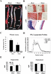
Figure 4. Quantitative analysis of atherosclerotic lesion size in aortas from cholesterol-fed mice with or without rosiglitazone treatment.
(A) Aortas from 20-week-old mice that express (LRP+) of lack (LRP) LRP in VSMC (n = 6 mice per group). Mice had been cholesterol-fed for 5 weeks in the absence (−Rosi) or presence (+Rosi) of rosiglitazone (GlaxoSmithKline, 25 mg/kg/day) before analysis. Aortae were stained en face with Sudan IV and arrows indicate lipid-laden (Sudan-positive) atherosclerotic lesions. Scale bar, 1.2 cm. (B) Histological analysis of thoracic aortas from animals cholesterol-fed in the absence or presence of rosiglitazone. Hematoxylin and eosin (a and b, LRP+; c and d, LRP), and trichrome staining (e and f, LRP) of longitudinal sections. Scale bar in a, 15 µm. (C) Quantitative analysis of atherosclerotic lesion size in aortas from cholesterol-fed LRP and control (LRP+) mice (n = 5 mice per group) with and without rosiglitazone treatment. Values are expressed as mean±s.e.m. *, p<0.05 for LRP treated versus untreated. (D) FPLC profile of plasma lipoproteins from untreated LRP (filled squares) and LRP+ (opened squares) and rosiglitazone treated LRP (filled triangles) and LRP+ (opened triangles). (E) Plasma triglycerides and (F) cholesterol from untreated and rosiglitazone treated LRP and LRP+ mice. Values are expressed as mean±S.E.M. (n = 10 mice per group).
Figure 5
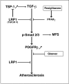
Figure 5. Control of TGFβ and PDGF signaling and protection of vascular wall integrity by LRP1.
Absence of LRP1 results in increased activation of TGFβ signaling. This is accompanied by disruption of elastic layers, tortuous aortas and increased fibrosis similar to what is observed in Marfan and Marfan-like syndromes in which the genes for fibrillin-1 or TGFβ receptors are defective. Loss of LRP1 expression leads to increased expression of PDGF receptors. LRP1 also controls PDGFRβ signaling and trafficking through an independent mechanism, and absence of LRP1 promotes VSMC proliferation and severe atherosclerosis. Rosiglitazone blocks TGFβ signaling upstream of PDGFR, which is inhibited by Gleevec. Both drugs are thus effective in reducing arterial wall thickening and atherosclerosis, which are induced by increased PDGFR signaling.

Similar articles

Cited by

References

    1. Boucher P, Gotthardt M, Li WP, Anderson RG, Herz J. LRP: role in vascular wall integrity and protection from atherosclerosis. Science. 2003;300:329–332. - PubMed
    1. Boucher P, Liu P, Gotthardt M, Hiesberger T, Anderson RG, et al. Platelet-derived growth factor mediates tyrosine phosphorylation of the cytoplasmic domain of the low Density lipoprotein receptor-related protein in caveolae. J Biol Chem. 2002;277:15507–15513. - PubMed
    1. Takayama Y, May P, Anderson RG, Herz J. Low density lipoprotein receptor-related protein 1 (LRP1) controls endocytosis and c-CBL-mediated ubiquitination of the platelet-derived growth factor receptor beta (PDGFR beta). J Biol Chem. 2005;280:18504–18510. - PubMed
    1. Boileau C, Jondeau G, Mizuguchi T, Matsumoto N. Molecular genetics of Marfan syndrome. Curr Opin Cardiol. 2005;20:194–200. - PubMed
    1. Dietz HC, Loeys B, Carta L, Ramirez F. Recent progress towards a molecular understanding of Marfan syndrome. Am J Med Genet C Semin Med Genet. 2005;139:4–9. - PubMed

Publication types

MeSH terms

Substances