. 2007 May 1;3(5):292-302.
doi: 10.7150/ijbs.3.292.

Human Maf1 negatively regulates RNA polymerase III transcription via the TFIIB family members Brf1 and Brf2

Affiliations

Human Maf1 negatively regulates RNA polymerase III transcription via the TFIIB family members Brf1 and Brf2

Janet Rollins et al. Int J Biol Sci. .

Abstract

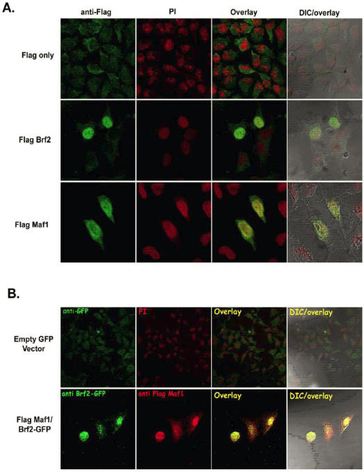
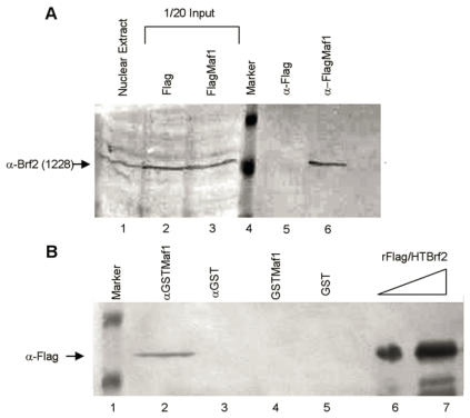
RNA polymerase III (RNA pol III) transcribes many of the small structural RNA molecules involved in processing and translation, thereby regulating the growth rate of a cell. Initiation of pol III transcription requires the evolutionarily conserved pol III initiation factor TFIIIB. TFIIIB is the molecular target of regulation by tumor suppressors, including p53, RB and the RB-related pocket proteins. However, our understanding of negative regulation of human TFIIIB-mediated transcription by other proteins is limited. In this study we characterize a RNA pol III luciferase assay and further demonstrate in vivo that a human homolog of yeast Maf1 represses RNA pol III transcription. Additionally, we show that Maf1 repression of RNA pol III transcription occurs via TFIIIB, specifically through the TFIIB family members Brf1 and Brf2.


Conflict of interest statement

Conflict of Interests: The authors have declared that no conflicts of interests exist.

Figures

Figure 1
Figure 1
RNA polymerase III transcription can be measured in vivo using a RNA pol III dual luciferase assay. (A) Transient transfection of HeLa cells with increasing concentrations of pGL3-U6 (100ng, 200ng, 300ng), pGL3-VAI (100ng, 200ng, 300ng) or empty pGL3 vector (300ng). (B) Luciferase activity comparison of the wild-type human U6 promoter (100ng) with a mutant human U6 promoter, lacking a functional TATA box and PSE (100ng). Cells were also transfected with empty pGL3 (100ng) as negative control. (C) Comparison of wild-type VAI (100ng) promoter luciferase activity with a mutant VAI construct lacking both the A and B box promoter elements of the VAI promoter (100ng). Cells were also transfected with empty pGL3 (100ng) as negative control. (D) HeLa cells transiently transfected with either empty pGL3 (50ng), wild-type pGL3-U6 (50ng), or a previously characterized mutant U6 lacking a functional TATA box and transcribed by RNA pol II, pGL3-mtTATAU6 (25ng) and treated with 0 μg/ml or 3 μg/ml α-amanitin. (E) HeLa cells transiently transfected with either empty pGL3 (100ng), wild-type pGL3-VAI (100ng), or a previously characterized mutant U6 lacking a functional TATA box and transcribed by RNA pol II, pGL3-mtTATAU6 (50ng) and treated with 0 μg/ml, 1 μg/ml or 2 μg/ml α-amanitin. (F) Transient transfection of pGL3 (100ng), pGL3-U6 (100ng), or pGL3-U6TTT (100ng) with a run of T residues inserted downstream of the human U6 promoter, but upstream of the the Kozak consensus translation initiation site of the luciferase gene in pGL3. All luciferase assay results expressed as relative light units (RLU): the average of the Photinus pyralis firefly activity observed divided by the average of the activity recorded from Renilla luciferase vector. Experiments were done in triplicate, repeated three times, representative experiments are depicted.
Figure 1
Figure 1
RNA polymerase III transcription can be measured in vivo using a RNA pol III dual luciferase assay. (A) Transient transfection of HeLa cells with increasing concentrations of pGL3-U6 (100ng, 200ng, 300ng), pGL3-VAI (100ng, 200ng, 300ng) or empty pGL3 vector (300ng). (B) Luciferase activity comparison of the wild-type human U6 promoter (100ng) with a mutant human U6 promoter, lacking a functional TATA box and PSE (100ng). Cells were also transfected with empty pGL3 (100ng) as negative control. (C) Comparison of wild-type VAI (100ng) promoter luciferase activity with a mutant VAI construct lacking both the A and B box promoter elements of the VAI promoter (100ng). Cells were also transfected with empty pGL3 (100ng) as negative control. (D) HeLa cells transiently transfected with either empty pGL3 (50ng), wild-type pGL3-U6 (50ng), or a previously characterized mutant U6 lacking a functional TATA box and transcribed by RNA pol II, pGL3-mtTATAU6 (25ng) and treated with 0 μg/ml or 3 μg/ml α-amanitin. (E) HeLa cells transiently transfected with either empty pGL3 (100ng), wild-type pGL3-VAI (100ng), or a previously characterized mutant U6 lacking a functional TATA box and transcribed by RNA pol II, pGL3-mtTATAU6 (50ng) and treated with 0 μg/ml, 1 μg/ml or 2 μg/ml α-amanitin. (F) Transient transfection of pGL3 (100ng), pGL3-U6 (100ng), or pGL3-U6TTT (100ng) with a run of T residues inserted downstream of the human U6 promoter, but upstream of the the Kozak consensus translation initiation site of the luciferase gene in pGL3. All luciferase assay results expressed as relative light units (RLU): the average of the Photinus pyralis firefly activity observed divided by the average of the activity recorded from Renilla luciferase vector. Experiments were done in triplicate, repeated three times, representative experiments are depicted.
Figure 2
Figure 2
Human Maf1 inhibits RNA polymerase III transcription from both gene internal and gene external promoters. (A) Anti-flag immunoblot of nuclear extract prepared from HeLa cells transiently transfected with: empty flag vector (lane 1), FlagMaf1 (lane 2), FlagBrf1 (lane 3), FlagBrf2 (lane 4). Arrows depict location of each protein as determined by anti-Flag antibody. Asterisks denote polypeptide doublet routinely observed in HeLa cells over-expressing FlagMaf1. (B) Same immunoblot from 2A reprobed using anti-actin antibody as loading control for transfections with FlagBrf1, FlagBrf2 and empty Flag vector. (C) Nuclear extract prepared from HeLa cells over-expressing FlagMaf1 treated with calf intestine phosphatase (CIP) for increasing amounts of time and immunoblotted with an anti-Flag antibody. (D) Transient transfection of HeLa cells with pGL3-VAI (100ng) (left panel) or pGL3-U6 (100ng) (right panel) with increasing amounts of FlagMaf1(50ng, 100ng, 200ng) and empty vectors Flag (200ng) and pGL3(100ng) as indicated by + sign.
Figure 3
Figure 3
Overexpression of the TFIIIB subunits Brf1 or Brf2 alleviates Maf1 induced repression of transcription from both gene internal and external promoters RNA polymerase III. (A) HeLa cells were transiently transfected with: pGL3 and Flag vector, or pGL3-VAI and Flag vector, pGL3-VAI and FlagMaf1, pGL3-VAI, FlagMaf1 and Brf1. (B) HeLa cells were transiently transfected with: pGL3 and Flag vector, or pGL3-U6 and Flag vector, pGL3-U6 and FlagMaf1, pGL3-U6, FlagMaf1 and Brf2.
Figure 4
Figure 4
Human Maf1 and Brf2 co-localize in HeLa cells. (A) HeLa cells transfected with empty Flag vector (left upper panel), FlagBrf2 (center left panel), or FlagMaf1 (lower left panel) and localization of proteins determined using an anti-Flag antibody denoted in green. Propidium iodide (PI), center left panels, identify the nucleus of HeLa cells. Overlay of cells stained with anti-Flag and PI demonstrate the nuclear localization of FlagMaf1 and FlagBrf2, right center panels. Right panels show the DIC and overlay of the transfected HeLa cells. (B) HeLa cells transfected with either empty Flag and GFP vectors or FlagMaf1 and Brf2GFP. Right panels demonstrate location of GFP signal (green), left center panels the Flag signal (red), overlay shows co-localization of FlagMaf1 and Brf2-GFP (yellow).
Figure 5
Figure 5
Maf1 directly associates with the TFIIB family member Brf2. (A) Nuclear extracts from HeLa cells transiently transfected with the empty Flag vector or FlagMaf1 were mixed with Flag agarose, subsequently immunoblotted with the gene specific anti-Brf2 antibody CSH 1228. Nuclear extract inputs: untransfected nuclear extract (lane 1) empty Flag vector (lane 2), and FlagMaf1 (lane 3). Immunoprecipitates: Flag vector (lane 5), and FlagMaf1 (lane 6). Arrow denotes migration location of endogenous Brf2 protein. (B) Recombinant Flag/HTBrf2 and GSTMaf1 were mixed with anti-glutathione agarose and immunoprecipitates were immunoblotted with an anti-Flag antibody. Increasing amounts of recombinant Flag/HTBrf2 (lanes 6 and 7), recombinant GST (lane 5), and recombinant GSTMaf1 (lane 4), immune complexes from anti-GST (lane 3) and anti-GSTMaf1 (lane 2) immunoprecipitations. Arrow denotes location of Flag/HT Brf2, as determined by anti-Flag antibody.

Similar articles

Cited by

References

    1. Roeder R.G. Nuclear RNA polymerases: role of general initiation factors and cofactors in eukaryotic transcription. Methods Enzymol. 1996;273:165–71. - PubMed
    1. Schramm L, Hernandez N. Recruitment of RNA polymerase III to its target promoters. Genes Dev. 2002;16(20):2593–620. - PubMed
    1. White R.J. RNA polymerases I and III, growth control and cancer. Nat Rev Mol Cell Biol. 2005;6(1):69–78. - PubMed
    1. Gabrielsen O.S, Sentenac A. RNA polymerase III (C) and its transcription factors. Trends Biochem Sci. 1991;16(11):412–6. - PubMed
    1. Desai N et al. Two steps in Maf1-dependent repression of transcription by RNA polymerase III. J Biol Chem. 2005;280(8):6455–62. - PubMed

Publication types

MeSH terms