Proteomics analysis of the expression of neurogranin in murine neuroblastoma (Neuro-2a) cells reveals its involvement for cell differentiation
- PMID: 17505539
- PMCID: PMC1865092
- DOI: 10.7150/ijbs.3.263
Proteomics analysis of the expression of neurogranin in murine neuroblastoma (Neuro-2a) cells reveals its involvement for cell differentiation
Abstract
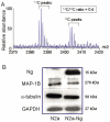
Neurogranin (Ng) is a neural-specific, calmodulin (CaM)-binding protein that is phosphorylated by protein kinase C (PKC). Although its biochemical property has been well characterized, the physiological function of Ng needs to be elucidated. In the present study, we performed proteomics analysis of the induced compositional changes due to the expression of Ng in murine neuroblastoma (Neuro-2a) cells using isotope coded affinity tags (ICAT) combined with 2-dimensional liquid chromatography/tandem mass spectrometry (2D-LC/MS/MS). We found that 40% of identified proteins were down-regulated and most of these proteins are microtubule components and associated proteins that mediated neurite outgrowth. Western blot experiments confirmed the expression of alpha-tubulin and microtubule- associated protein 1B (MAP 1B) was dramatically reduced in Neuro-2a-Ng cells compared to control. Cell morphology of Neuro-2a-Ng showed far less neurites than the control. Serum deprivation induced the extension of only one or two long neurites per cell in Neuro-2a-Ng, contrasting to the extension of multiple neurites per control cell. Ng may be linked to neurite formation by affecting expression of several microtubule related proteins. Furthermore, the PKC activator (PMA) induced an enhanced ERK1/2 activity in the cells that expressed Ng. The mutation of Ng at S36A caused sustained increase of ERK1/2 activity, whereas the ERK1/2 activity in mutation at I33Q showed no difference compared to wild type Ng, suggesting the phosphorylation of Ng but not the CaM /Ng interaction plays an important role in ERK activation. Ng may be involved in neuronal growth and differentiation via PKC and ERK1/2 signaling pathways.
Conflict of interest statement
CONFLICT OF INTEREST: The authors have declared that there is no conflict of interest.
Figures


Similar articles
-
Characterization of transcriptional regulation of neurogranin by nitric oxide and the role of neurogranin in SNP-induced cell death: implication of neurogranin in an increased neuronal susceptibility to oxidative stress.Int J Biol Sci. 2007 Feb 23;3(4):212-24. doi: 10.7150/ijbs.3.212. Int J Biol Sci. 2007. PMID: 17389928 Free PMC article.
-
Ionizing radiation induces neuronal differentiation of Neuro-2a cells via PI3-kinase and p53-dependent pathways.Int J Radiat Biol. 2015 Jul;91(7):585-95. doi: 10.3109/09553002.2015.1029595. Epub 2015 May 20. Int J Radiat Biol. 2015. PMID: 25912236
-
A casein kinase II-related activity is involved in phosphorylation of microtubule-associated protein MAP-1B during neuroblastoma cell differentiation.J Cell Biol. 1988 Jun;106(6):2057-65. doi: 10.1083/jcb.106.6.2057. J Cell Biol. 1988. PMID: 3164313 Free PMC article.
-
Neurogranin, a link between calcium/calmodulin and protein kinase C signaling in synaptic plasticity.IUBMB Life. 2010 Aug;62(8):597-606. doi: 10.1002/iub.357. IUBMB Life. 2010. PMID: 20665622 Review.
-
Quantitative neuroproteomics: classical and novel tools for studying neural differentiation and function.Stem Cell Rev Rep. 2011 Mar;7(1):77-93. doi: 10.1007/s12015-010-9136-3. Stem Cell Rev Rep. 2011. PMID: 20352529 Review.
Cited by
-
Structural basis for the interaction of unstructured neuron specific substrates neuromodulin and neurogranin with Calmodulin.Sci Rep. 2013;3:1392. doi: 10.1038/srep01392. Sci Rep. 2013. PMID: 23462742 Free PMC article.
-
Intrahippocampal injection of a lentiviral vector expressing neurogranin enhances cognitive function in 5XFAD mice.Exp Mol Med. 2018 Mar 23;50(3):e461. doi: 10.1038/emm.2017.302. Exp Mol Med. 2018. PMID: 29568074 Free PMC article.
-
Neuronal NOS Induces Neuronal Differentiation Through a PKCα-Dependent GSK3β Inactivation Pathway in Hippocampal Neural Progenitor Cells.Mol Neurobiol. 2017 Sep;54(7):5646-5656. doi: 10.1007/s12035-016-0110-1. Epub 2016 Sep 13. Mol Neurobiol. 2017. PMID: 27624386
-
Zinc regulation of transcriptional activity during retinoic acid-induced neuronal differentiation.J Nutr Biochem. 2013 Nov;24(11):1940-4. doi: 10.1016/j.jnutbio.2013.06.002. Epub 2013 Sep 9. J Nutr Biochem. 2013. PMID: 24029070 Free PMC article.
-
Expression and secretion of synaptic proteins during stem cell differentiation to cortical neurons.Neurochem Int. 2018 Dec;121:38-49. doi: 10.1016/j.neuint.2018.10.014. Epub 2018 Oct 18. Neurochem Int. 2018. PMID: 30342961 Free PMC article.
References
-
- Baudier J, Deloulme JC, Van Dorsselaer A, Black D, Matthes HW. Purification and characterization of a brain-specific protein kinase C substrate, neurogranin (p17). Identification of a consensus amino acid sequence between neurogranin and neuromodulin (GAP43) that corresponds to the protein kinase C phosphorylation site and the calmodulin-binding domain. J Biol Chem. 1991;266:229–237. - PubMed
-
- Sheu FS, Mahoney CW, Seki K, Huang KP. Nitric oxide modification of rat brain neurogranin affects its phosphorylation by protein kinase C and affinity for calmodulin. J Biol Chem. 1996;271:22407–22413. - PubMed
-
- Cohen RW, Margulies JE, Coulter PM 2nd, Watson JB. Functional consequences of expression of the neuron-specific, protein kinase C substrate RC3 (neurogranin) in Xenopus oocytes. Brain Res. 1993;627:147–152. - PubMed
-
- Yang HM, Lee PH, Lim TM, Sheu FS. Neurogranin expression in stably transfected N2A cell line affects cytosolic calcium level by nitric oxide stimulation. Brain Res Mol Brain Res. 2004;129:171–178. - PubMed
Publication types
MeSH terms
Substances
LinkOut - more resources
Full Text Sources
Other Literature Sources
Miscellaneous

