Review
. 2007:76:267-94.
doi: 10.1146/annurev.biochem.75.103004.142811.

Synapses: sites of cell recognition, adhesion, and functional specification

Affiliations
Review

Synapses: sites of cell recognition, adhesion, and functional specification

Soichiro Yamada et al. Annu Rev Biochem. 2007.

Abstract

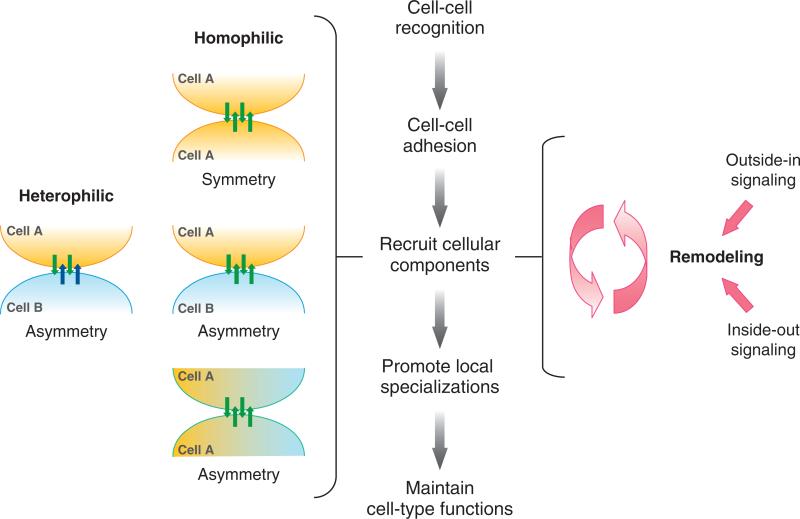
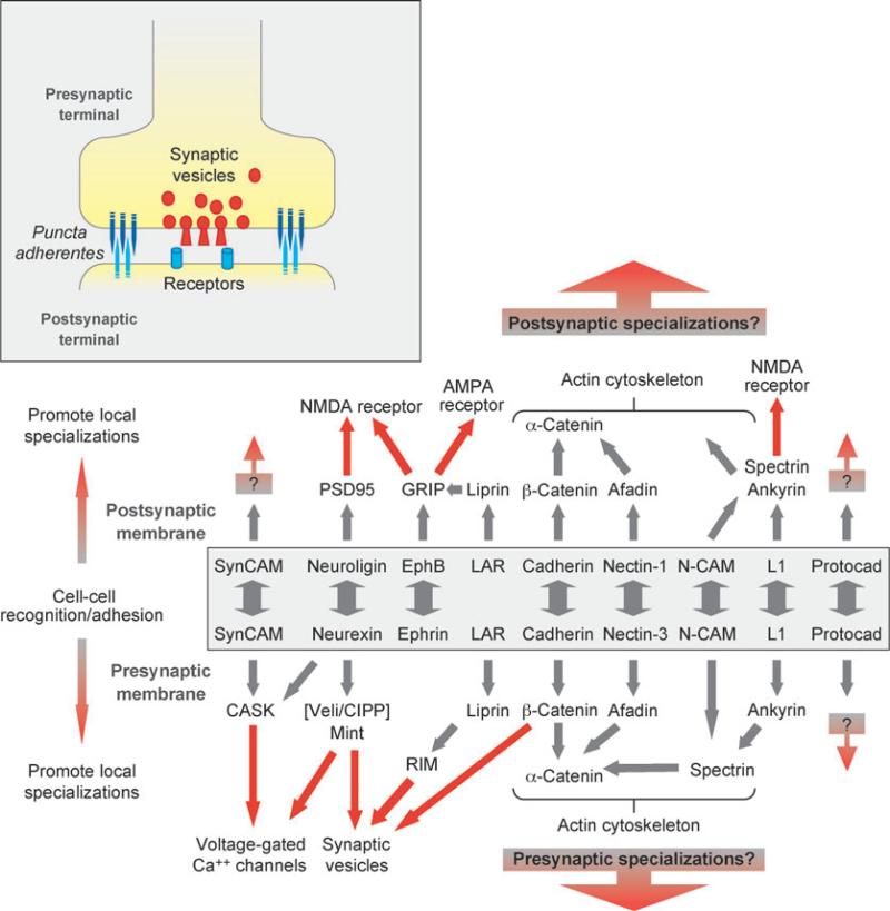
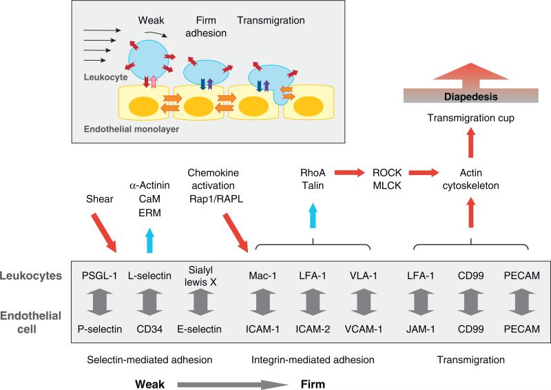
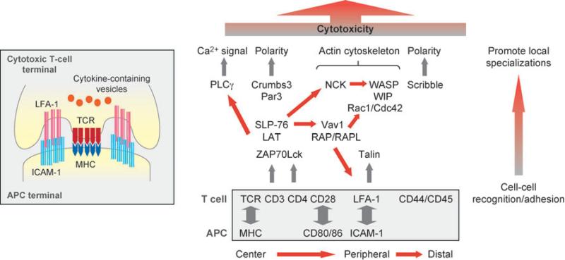
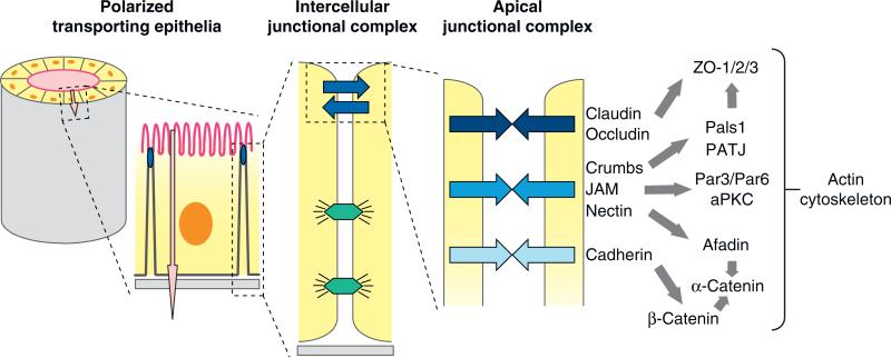
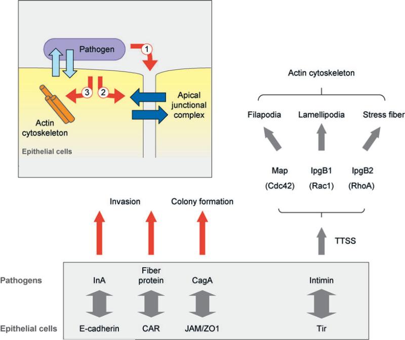
Synapses are specialized adhesive contacts characteristic of many types of cell-cell interactions involving neurons, immune cells, epithelial cells, and even pathogens and host cells. Cell-cell adhesion is mediated by structurally diverse classes of cell-surface glycoproteins, which form homophilic or heterophilic interactions across the intercellular space. Adhesion proteins bind to a cytoplasmic network of scaffolding proteins, regulators of the actin cytoskeleton, and signal transduction pathways that control the structural and functional organization of synapses. The themes of this review are to compare the organization of synapses in different cell types and to understand how different classes of cell adhesion proteins and cytoplasmic protein networks specify the assembly of functionally distinct synapses in different cell contexts.


Figures

Figure 1
Figure 1
Schematic representation of sequential events following cell recognition and adhesion that promote local specialization of the synapse. (left) Heterophilic adhesion between different cells (A, B), expressing different adhesion proteins (blue and green arrows), leads to the formation of an asymmetric synapse; homophilic adhesion between the same cell types (A, A), expressing the same adhesion proteins (green arrows), leads to formation of a symmetric synapse and either a symmetric organization of cells (top), an asymmetric organization of the same cells (A, A) in the apicobasal cell axis (middle), or an asymmetric organization between two different cells (A, B). (right) The organization of adhesion proteins and cytoplasmic signaling complexes can be remodeled by either outside-in or inside-out signaling.
Figure 2
Figure 2
Schematic representations of the neuronal synapse (inset) and protein interactions at the pre- and postsynaptic membranes. Different cell adhesion proteins form homophilic or heterophilic adhesions (boxed) and interact with downstream protein networks that describe functional specification of the presynaptic (recruit Ca2+ channels, synaptic vesicles) and postsynaptic membranes (recruit neurotransmitter receptors). Abbreviations: AMPA, α-amino-5-hydroxy-3-methyl-4-isoxazole propionic acid; CASK, calcium/calmodulin-dependent serine protein kinase; CIPP, channel-interacting PDZ domain protein; GRIP, glutamate receptor-interacting protein; LAR, leukocyte common antigen-related protein; Mint, Munc-18-interacting protein; N-CAM, neural cell adhesion molecule; NMDA, N-methyl-D-aspartic acid; PSD95, postsynaptic density 95; RIM, Rab3-interacting molecule; Veli, vertebrate LIN-7.
Figure 3
Figure 3
Schematic representation of leukocyte-endothelial cell interactions during extravasation (inset). Selectin-mediated adhesion (red/pink arrows) initiates leukocyte rolling, and integrin-mediated adhesion (blue/purple arrows) stabilizes leukocyte adhesion to endothelial cells. Endothelial junction proteins (orange arrows) mediate leukocyte transmigration. Adhesion proteins (box) and protein networks assembled at the synapse between rolling leukocytes and endothelial cells (selectin- and integrin-mediated adhesion) as well as during leukocyte transmigration (JAM-1/LFA-1, CD99, and PECAM). Abbreviations: CaM, calmodulin; ERM, ezrin/radixin/moesin; MLCK, myosin light chain kinase; RAPL, regulator of adhesion and cell polarization enriched in lymphoid tissues; ROCK, Rho kinase.
Figure 4
Figure 4
Schematic representations of the immunologic synapse between T cells and antigen-presenting cells (APCs) (inset), adhesion proteins (boxed), and protein networks assembled at the synapse that remodel the T cell and initiate the immune response from the T cell. Abbreviations: ICAM-1, intercellular adhesion molecule 1; LAT, linker for activation of T cells; LFA-1, lymphocyte function-associated antigen 1; MHC, major histocompatibility complex; Par3, partition defective 3; PLCγ, phospholipase C γ; SLP-76, Src homology 2 domain-containing leukocyte-specific phosphoprotein of 76 kDa; TCR, T-cell receptor; WASP, Wiskott-Aldrich syndrome protein; WIP, WASP-interacting protein; ZAP70, zeta-chain-associated protein 70.
Figure 5
Figure 5
Schematic representations of the epithelial synapse. Polarized transporting epithelia comprise a closed monolayer of cells that surround a fluid-filed space (lumen) and vectorially transport ions and solutes (pink arrow) between the luminal space and the serosa; the plasma membrane domains facing the lumen space (apical) and serosa (basal lateral) are structurally and functionally different. Intercellular junctional complexes regulate cell-cell adhesion and the paracellular pathway (blue arrows) as well as maintain the structural integrity of the epithelium (desmosomes, lower junctions). The apical junctional complex comprises different cell adhesion proteins and downstream protein networks, and it is located at the boundary between the apical and basal-lateral membranes. Abbreviations: aPKC, atypical protein kinase C; JAM, junction-associated molecule; Pals1, protein-associated with Lin-7; Par3/Par6, partition defective 3/6; PATJ, Pals1-associated tight junction protein.
Figure 6
Figure 6
Schematic representations of novel synapses formed between pathogens and host cells. Pathogens form synapses on the luminal surface of epithelial cells (inset) and modify the host cell synapse from the outside (1) or inside (2), or through reorganization of the actin cytoskeleton (3). Pathogen surface proteins bind host cell adhesion proteins (left) or a pathogen protein inserted by a type III secretion system (TTSS) (right), and hijack host protein networks to modify the actin cytoskeleton and membrane dynamics. Abbreviations: CagA, cytotoxin-associated gene 1; CAR, Coxsackie virus and adenovirus receptor; InlA, internalin A; JAM, junction-associated molecule; Map, mitochondrial-associated protein; Tir, translocated intimin receptor; ZO1, zonula occludens-1.

References

    1. Bennett MR. Brain Res. Bull. 1999;50:95–118. - PubMed
    1. Cunningham BA, Hemperly JJ, Murray BA, Prediger EA, Brackenbury R, Edelman GM. Science. 1987;236:799–806. - PubMed
    1. Bruses JL, Rutishauser U. Biochimie. 2001;83:635–43. - PubMed
    1. Cremer H, Lange R, Christoph A, Plomann M, Vopper G, et al. Nature. 1994;367:455–59. - PubMed
    1. Hu H, Tomasiewicz H, Magnuson T, Rutishauser U. Neuron. 1996;16:735–43. - PubMed

Publication types

MeSH terms