Review
. 2007:30:235-58.
doi: 10.1146/annurev.neuro.30.051606.094345.

Neuron-glial interactions in blood-brain barrier formation

Affiliations
Review

Neuron-glial interactions in blood-brain barrier formation

Swati Banerjee et al. Annu Rev Neurosci. 2007.

Abstract

The blood brain barrier (BBB) evolved to preserve the microenvironment of the highly excitable neuronal cells to allow for action potential generation and propagation. Intricate molecular interactions between two main cell types, the neurons and the glial cells, form the underlying basis of the critical functioning of the nervous system across species. In invertebrates, interactions between neurons and glial cells are central in establishing a functional BBB. However, in vertebrates, the BBB formation and function is coordinated by interactions between neurons, glial cells, and endothelial cells. Here we review the neuron-glial interaction-based blood barriers in invertebrates and vertebrates and provide an evolutionary perspective as to how a glial-barrier system in invertebrates evolved into an endothelial barrier system. We also summarize the clinical relevance of the BBB as this protective barrier becomes disadvantageous in the pharmacological treatment of various neurological disorders.


Figures

Figure 1
Figure 1
Distribution and migration of glial cells in the Drosophila embryos. (a–c) A portion of a stage 12 embryo immunostained against a motor neuron marker, anti-FAS II (green), and a glial marker, anti-REPO (red). The glial cells are in close proximity of the motor neurons in the neurectoderm, which produces both neuroblasts and glioblasts. (d–f) A portion of a stage 14 embryo showing migration of the glia toward the periphery. The FAS II positive axons have exited the CNS. The glial cells are lined along the axons and are still migrating to their final destination. These close interactions between neurons and glial cells eventually allow the ensheathment of neurons and establishment of the BBB.
Figure 2
Figure 2
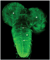
Perineurial glial cells and the BBB. Immunolocalization of Nrx IV in the larval ventral nerve cord and brain lobes of the CNS shows that the protein is expressed in midline glia (arrows) as well as at the edges of the perineurial glial cells required for the maintenance of the BBB in third instar larvae. These are very large cells that ensheath the entire CNS. Note the giant cells surrounding the brain lobes and also the nerve cord (asterisks). Anterior is up.
Figure 3
Figure 3
Breakdown of the BBB. (a) A wild-type stage 16 Drosophila embryo injected with a 10-KDa rhodamine-dextran dye shows the presence of functional SJs because the dye does not penetrate any of the organs. (b) neurexin IV (nrx–/–) and (c) neuroglian (nrg–/–) mutant embryos, which lack SJs, show a breakdown of the BBB as the dye penetrates the CNS (arrows) and other organs.
Figure 4
Figure 4
Distribution and final position of peripheral glia. (a) A portion of a stage 16 wild-type Drosophila embryo stained with a motor axon marker, Fasciclin II (FAS II, green) and a glial marker REPO (red). The axons have completed their exit from the CNS and have grown toward the periphery. The white dotted line indicates a presumptive boundary that separates the CNS from the periphery. Some of the glial cells that can be clearly identified in this focal plane are EG (exit glia) and PG (peripheral glia). (b) A portion of a stage 16 wild-type embryo stained with sensory neuron marker 22C10 (green) and REPO (red). The axons from the sensory neuronal clusters (dorsal, D; lateral chordotonal cluster, LCH; ventral prime, V′ and ventral, V) have made their way into CNS. The glial cells highlighted in (a) are also marked here. The sensory and motor axons in a segment use the same paths and are always insulated together. This ensheathment ensures a functional BNB in PNS. (For further details, see Klambt & Goodman 1991.)
Figure 5
Figure 5
Disruption in the chordotonal organ (CO) morphology and BNB function in nrx IV, cont, and nrg mutants. (a) Schematic of a CO showing the various cell types. Cap cells (cc, green), scolopale (sc, red), and ligament (lig) are the three glial cell types. The neuron (neu, blue) has a rootlet (r) and its dendrite (d) projects into the lumen (lu) of the scolopale. The cc and the lig cell attachment sites of the CO to the epidermis (ep) are also shown. The presence of extensive SJs is apparent between the cap and scolopale cells, thus providing a functional BNB. (b) Wild-type CO triple stained with antiβ3tubulin (green) marking the cap cells, anti-Crb (red) marking the lumen of the scolopale, and anti22C10 (blue) marking the sensory neuron. (c–h) Wild-type COs (c, e, and g) stained with anti-CRB (red) and anti22C10 (blue) in combination with anti-Nrx IV (c, d), anti-Cont (e, f), and anti-Nrg (g–h) show a fusiform shape of the scolopales in the CO cluster. nrx IV (d), cont (f), and nrg (h) mutants as evident from the lack of staining of their respective antibodies show a defective morphology and a disarrayed organization of the cluster. (i–l) Dye exclusion assays performed on the wild-type embryos (i), nrx IV (j), cont (k) and nrg (l) mutant embryos. Confocal images after dye injection of the regions of the peripheral nervous system at the level of the COs. Wild-type embryos (i) excluded the dye from the COs even after 30 min of injection, indicating that a functional BNB is present. Under identical conditions nrx IV (j), cont (k), and nrg (l) mutant embryos failed to exclude the dye from the COs. Confocal images showed dye penetration into COs within 15 min after injection, indicating that the BNB has broken down in these mutants. Printed with permission from Banerjee et al. (2006a); copyright 2006 by the Society for Neuroscience.
Figure 6
Figure 6
Schematic of the neurovascular unit. (a) A cross-section through a brain capillary shows adjacent endothelial cells connected by TJs that establish the BBB. The endothelial cell layer is surrounded by the basal lamina that separates the endothelium from the pericytes, astrocytes, and neurons. (b) A longitudinal section through a portion of a brain capillary reveals the presence of adjacent endothelial cells connected by TJs. Pericytes are present within the basal lamina in close proximity to the endothelial cells, whereas astrocytic endfeet are on the outer surface of the basal lamina. Microglia, nerve fibers, and neuromuscular synapses are found in the perivascular space. Panel b has been modified with permission from Abbott 2005; copyright 2005 by Springer Science and Business Media.

References

    1. Abbott NJ. Comparative physiology of the blood-brain barrier. In: Bradbury MWB, editor. Physiology and Pharmacology of the Blood-Brain-Barrier. Berlin: Springer-Verlag; 1992. pp. 371–96.
    1. Abbott NJ. Inflammatory mediators and modulation of blood-brain barrier permeability. Cell Mol Neurobiol. 2000;20:131–47. - PMC - PubMed
    1. Abbott NJ. Astrocyte-endothelial interactions and blood-brain barrier permeability. J Anat. 2002;200:629–38. - PMC - PubMed
    1. Abbott NJ. Dynamics of CNS barriers: evolution, differentiation, and modulation. Cell Mol Neurobiol. 2005;25:5–23. - PMC - PubMed
    1. Abbott NJ, Bundgaard M, Cserr HF. Tightness of the blood-brain barrier and evidence for brain interstitial fluid flow in the cuttlefish, Sepia officinalis. J Physiol. 1985;368:213–26. - PMC - PubMed

Publication types

LinkOut - more resources