Review
. 2007:23:345-73.
doi: 10.1146/annurev.cellbio.23.090506.123249.

Cilia and developmental signaling

Affiliations
Review

Cilia and developmental signaling

Jonathan T Eggenschwiler et al. Annu Rev Cell Dev Biol. 2007.

Abstract

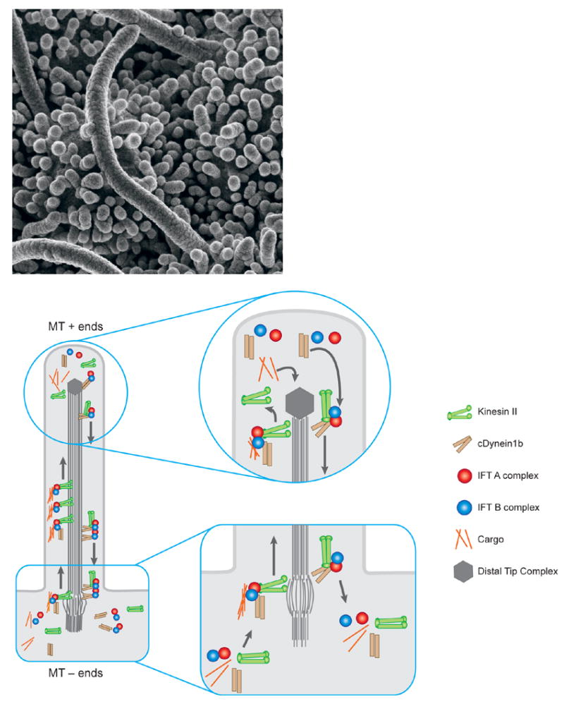
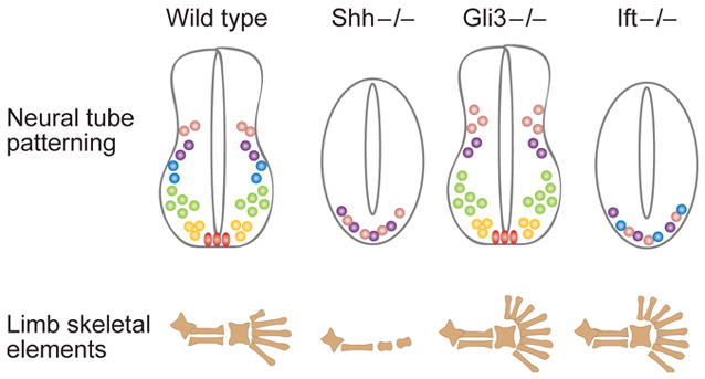
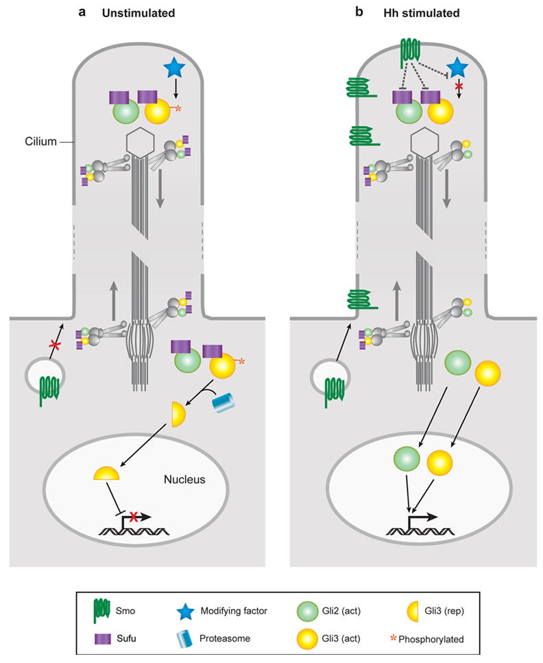
Recent studies have revealed unexpected connections between the mammalian Hedgehog (Hh) signal transduction pathway and the primary cilium, a microtubule-based organelle that protrudes from the surface of most vertebrate cells. Intraflagellar transport proteins, which are required for the construction of cilia, are essential for all responses to mammalian Hh proteins, and proteins required for Hh signal transduction are enriched in primary cilia. The phenotypes of different mouse mutants that affect ciliary proteins suggest that cilia may act as processive machines that organize sequential steps in the Hh signal transduction pathway. Cilia on vertebrate cells are likely to be important in additional developmental signaling pathways and are required for PDGF receptor alpha signaling in cultured fibroblasts. Cilia are not essential for either canonical or noncanonical Wnt signaling, although cell-type-specific modulation of cilia components may link cilia and Wnt signaling in some tissues. Because ciliogenesis in invertebrates is limited to a very small number of specialized cell types, the role of cilia in developmental signaling pathways is likely a uniquely vertebrate phenomenon.


Figures

Figure 1
Figure 1. Cilia and IFT
a. Cilia on the node of the e8.0 mouse embryo. These cilia are roughly 3–4 microns long, relatively longer than typical primary cilia. b. A schematic of the process of Intraflagellar Transport (IFT). Construction and maintenance of cilia and flagella depends on a steady source of new components, which are assembled at the distal tip complex (DTC, at the plus ends of the microtubules). Transport to the distal tip involves assembly of the anterograde IFT platform on transition fibers at the base of the growing cilium and anterograde movement powered by the kinesin II motor. Though still mechanistically unclear, events at the DTC are thought to involve disassembly of the anterograde transport machinery, assembly of structural components into the growing axoneme, and assembly/engagement of the retrograde transport apparatus. IFT machinery and residual structural cargo are recycled by retrograde transport from the tip to the base of the cilium powered by the cytoplasmic dynein motor. At the base, the retrograde apparatus is disassembled, potentially allowing IFT components to be reused.
Figure 2
Figure 2. Tissue-specific effects of IFT mutants depend on the importance of Gli activator and Gli repressor in the tissue
All Hedgehog signaling in the mouse depends on IFT proteins. Mammalian Hedgehog signaling has two outputs: production of Gli activator and prevention of processing of Gli3 repressor. In the neural tube, Gli activators play a central role in neural patterning, so loss of IFT in that tissue has a phenotype similar to that of Shh mutants. The IFT mutant neural tube phenotype is not identical to that of Shh mutants because Gli3 repressor (which is present and active in Shh mutants) is absent in IFT mutants; thus there is a low level of pathway activation. In the limb, Gli3 repressor plays a central role in digit patterning, and the phenotype caused by loss of IFT is similar to that caused by loss of Gli3. Nevertheless, analysis of molecular targets shows that Gli activators also fail to function in the IFT mutant limb.
Figure 3
Figure 3
A model for Hedgehog signal transduction within the cilium. In the absence of stimulation, Gli proteins and Su(fu) are transported to and from the tip of the cilium. Within the cilium, Gli3 protein is phosphorylated, targeting it for cleavage into the transcriptional repressor form once it encounters the proteasome at the cilium base. Gli proteins are maintained in an inactive state by Su(fu) and other components. Upon stimulation by Hh, activated Smoothened in vesicles is targeted to the cilium membrane where it can spread to the cilium tip. Here, Smo antagonizes downstream inhibitors such as Su(fu) and blocks phosphorylation of Gli3. Released from inhibition, activated forms of Gli proteins are transported to the cell body and ultimately enter the nucleus where they stimulate the expression of Hh target genes.
Figure 4
Figure 4
Canonical Wnt signaling in the mouse embryo is normal in Ift172 mutant embryos. The Topgal transgene contains multimerized Lef/Tcf binding sites driving the expression of β-galactosidase and acts as a faithful reporter for sites of endogenous Wnt signaling (DasGupta et al, 1999). a. A wild type e10.5 embryo that carries a copy of the Topgal reporter. β-galactosidase is expressed in many distinct sites where canonical Wnt signaling is active at this stage of development. B. Homozygous Ift172 (wim) embryos that carry a copy of the Topgal reporter show the characteristic morphology of Ift mutants; the most obvious phenotype is an open neural tube that lacks the normal ventral groove of the neural tissue. The Topgal reporter is expressed in the same set of cell types in the mutant as in the wild type, and at indistinguishable levels.

References

    1. Adler PN. Planar signaling and morphogenesis in Drosophila. Dev Cell. 2002;2(5):525–35. - PubMed
    1. Afzelius BA. A human syndrome caused by immotile cilia. Science. 1976;193(4250):317–9. - PubMed
    1. Afzelius BA. Cilia-related diseases. J Pathol. 2004;204(4):470–7. - PMC - PubMed
    1. Albrecht-Buehler G. Phagokinetic tracks of 3T3 cells: parallels between the orientation of track segments and of cellular structures which contain actin or tubulin. Cell. 1977;12(2):333–9. - PubMed
    1. Ansley SJ, Badano JL, Blacque OE, Hill J, Hoskins BE, et al. Basal body dysfunction is a likely cause of pleiotropic Bardet-Biedl syndrome. Nature. 2003;425(6958):628–33. - PubMed

Publication types

Substances

LinkOut - more resources