. 2007 May 16:7:30.
doi: 10.1186/1472-6807-7-30.

The crystal structure of Escherichia coli TdcF, a member of the highly conserved YjgF/YER057c/UK114 family

Affiliations

The crystal structure of Escherichia coli TdcF, a member of the highly conserved YjgF/YER057c/UK114 family

Julia D Burman et al. BMC Struct Biol. .

Abstract

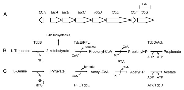
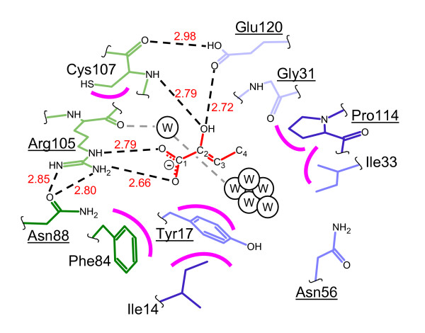
Background: The YjgF/YER057c/UK114 family of proteins is widespread in nature, but has as yet no clearly defined biological role. Members of the family exist as homotrimers and are characterised by intersubunit clefts that are delineated by well-conserved residues; these sites are likely to be of functional significance, yet catalytic activity has never been detected for any member of this family. The gene encoding the TdcF protein of E. coli, a YjgF/YER057c/UK114 family member, resides in an operon that strongly suggests a role in the metabolism of 2-ketobutyrate for this protein.

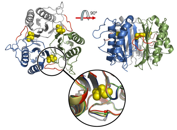
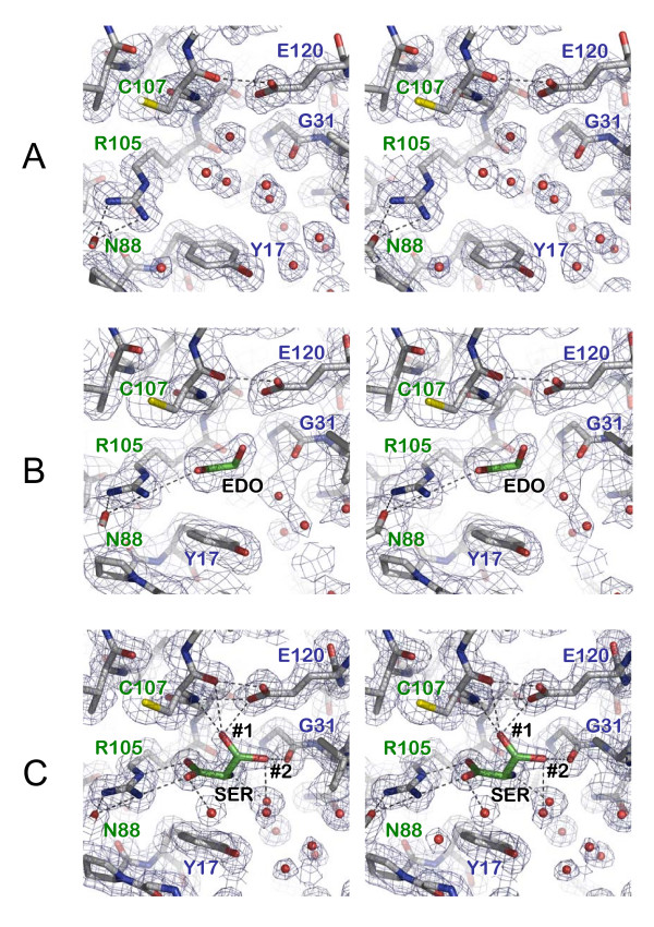
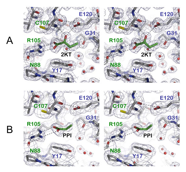
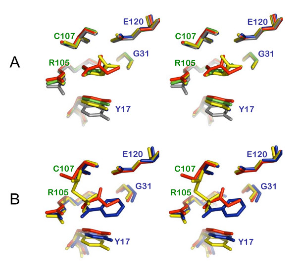
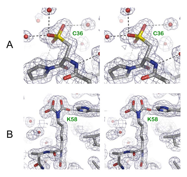
Results: We have determined the crystal structure of E. coli TdcF by molecular replacement to a maximum resolution of 1.6 A. Structures are also presented of TdcF complexed with a variety of ligands.

Conclusion: The TdcF structure closely resembles those of all YjgF/YER057c/UK114 family members determined thus far. It has the trimeric quaternary structure and intersubunit cavities characteristic of this family of proteins. We show that TdcF is capable of binding several low molecular weight metabolites bearing a carboxylate group, although the interaction with 2-ketobutyrate appears to be the most well defined. These observations may be indicative of a role for TdcF in sensing this potentially toxic metabolite.


Figures

Figure 1
Figure 1
The tdc operon and the pathways for degradation of L-threonine and L-serine. Schematic representation of the organisation of the tdc operon on the chromosome of E. coli and the function of the respective gene products. (A) shows the tdc operon [15], where the tdcA and tdcR gene products are transcriptional regulators, while the tdcC gene product is L-threonine/L-serine permease [30]. Also shown are the metabolic pathways for the anaerobic degradation of (B) L-threonine and (C) L-serine, where: Ack = acetate kinase; PFL = pyruvate formate-lyase; PTA = phosphotransacetylase.
Figure 2
Figure 2
The overall structure of E. coli TdcF. Ribbon representation of the E. coli TdcF structure. Shown is the 2-ketobutyrate-bound form with the individual subunits coloured green, blue and grey; the 2-ketobutyrate ligands are shown in space-filling representation in yellow. Part of the loop between β1 and β2 that moves depending on the status of the ligand-binding pocket is shown in red for each subunit. The inset shows the extent of this movement in the various states, where: grey = empty site; blue = ethylene glycol-bound; yellow = serine-bound; green = propionate-bound; red = 2-ketobutyrate-bound. The small spheres indicate the variation in the position of the Cα atom of Ile-14, being the residue that is displaced the most (maximum displacement 4.2Å). It is notable that the loop is poorly ordered in the empty site and best resolved in the in the 2-ketobutyrate-bound site, where Ile-14 makes non-bonding interactions with the ligand. Figure generated using PyMOL [31].
Figure 3
Figure 3
The ligand-binding pocket of TdcF in the empty, ethylene glycol-bound and serine-bound states. Stereoviews showing 2mFobs - dFcalc electron density maps contoured at approximately 1 sigma superposed on TdcF binding pockets in the following states: (A) empty (1.6 Å resolution); (B) with ethylene glycol bound (2.35 Å resolution); (C) with serine bound (1.6 Å resolution), where #1 and #2 denote alternate conformers for the Oγ. Important hydrogen bonds are shown as dashed lines. Residues from different subunits are labelled green and blue, respectively. In all cases, the side-chain of Arg-105 makes a bi-dentate interaction with the Oδ1 of Asn-88, which is just visible in the left foreground; and Oε1 of Glu-120 makes an inter-subunit hydrogen bond with the carbonyl oxygen of Cys-107. Throughout this figure, the view is similar to that seen in the inset of Figure 2. Figure generated using PyMOL [31].
Figure 4
Figure 4
The ligand-binding pocket of TdcF in the ketobutyrate-bound and propionate-bound states. Stereoviews, prepared as for Figure 3, showing TdcF binding pockets in the following states: (A) with 2-ketobutyrate bound (1.6 Å resolution); (B) with propionate bound (2.45 Å resolution).
Figure 5
Figure 5
Comparisons of the TdcF ligand-binding pocket. Stereoviews depicting comparisons of the TdcF ligand-binding pocket. (A) shows a superposition of all the states displayed in Figures 3 and 4, where: grey = empty site; blue = ethylene glycol-bound (the ethylene glycol itself is mainly occluded by other ligands in this view); yellow = serine-bound; red = 2-ketobutyrate-bound; green = propionate-bound. (B) shows a superposition of the ligand-binding site from TdcF with 2-ketobutyrate bound in red, onto the site from hp14.5 with a benzoic acid molecule bound in blue (primary site only)(PDB accession code 1ONI), and the equivalent site in YjgF in yellow (PDB accession code 1QU9). In the latter, two conformers are seen for Cys-107, the major one being covalently modified and protruding into the ligand-binding pocket. Throughout, the view is identical to that seen in Figures 3 and 4. Figure generated using PyMOL [31].
Figure 6
Figure 6
Covalent modifications of TdcF. Stereoviews showing covalent modifications of TdcF. In both cases, 1.6 Å resolution 2mFobs - dFcalc electron density maps are shown contoured at approximately 1 sigma. (A) a fully oxidised cysteine (cysteine sulfonic acid) is observed at position 36 in each subunit of all structures; (B) a carboxylated lysine in two alternative conformations that is visible in only the A chain of the 2-ketobutyrate-bound structure. Figure generated using PyMOL [31].
Figure 7
Figure 7
The binding of 2-ketobutyrate to TdcF. Schematic representation of the binding of 2-ketobutyrate to TdcF. 2-ketobutyrate is shown in red, whilst residues from one subunit are in green and those from the other subunit are in blue; darker colours indicate residues in the foreground and paler colours are used for those residues in the background. Underlining indicates strictly conserved residues in the YjgF/YER057c/UK114 family [8]. Important hydrogen bonds are shown as dashed lines and the curved magenta lines indicate non-bonding interactions with the ligand. The numbers in red give average hydrogen bond lengths in Å. Water molecules are indicated by the letter "W". The cluster of water molecules to the right of the ligand is in direct contact with bulk solvent. The view is similar to that seen in Figures 3–5.

Similar articles

Cited by

References

    1. Leitner-Dagan Y, Ovadis M, Zuker A, Shklarman E, Ohad I, Tzfira T, Vainstein A. CHRD, a plant member of the evolutionarily conserved YjgF family, influences photosynthesis and chromoplastogenesis. Planta. 2006;225:89–102. doi: 10.1007/s00425-006-0332-y. - DOI - PubMed
    1. Enos-Berlage JL, Langendorf MJ, Downs DM. Complex metabolic phenotypes caused by a mutation in yjgF, encoding a member of the highly conserved YER057c/YjgF family of proteins. J Bacteriol. 1998;180:6519–6528. - PMC - PubMed
    1. Schmitz G, Downs DM. Reduced transaminase B (IlvE) activity caused by the lack of yjgF is dependent on the status of threonine deaminase (IlvA) in Salmonella enterica serovar Typhimurium. J Bacteriol. 2004;186:803–810. doi: 10.1128/JB.186.3.803-810.2004. - DOI - PMC - PubMed
    1. Kim JM, Yoshikawa H, Shirahige K. A member of the YER057c/yjgf/Uk114 family links isoleucine biosynthesis and intact mitochondria maintenance in Saccharomyces cerevisiae. Genes Cells. 2001;6:507–517. doi: 10.1046/j.1365-2443.2001.00443.x. - DOI - PubMed
    1. Goupil-Feuillerat N, Cocaign-Bousquet M, Godon JJ, Ehrlich SD, Renault P. Dual role of alpha-acetolactate decarboxylase in Lactococcus lactis subsp. lactis. J Bacteriol. 1997;179:6285–6293. - PMC - PubMed

Publication types

MeSH terms

LinkOut - more resources