. 2007;8(5):R85.
doi: 10.1186/gb-2007-8-5-r85.

A global view of gene expression in lithium and zinc treated sea urchin embryos: new components of gene regulatory networks

Affiliations

A global view of gene expression in lithium and zinc treated sea urchin embryos: new components of gene regulatory networks

Albert J Poustka et al. Genome Biol. 2007.

Abstract

Background: The genome of the sea urchin Strongylocentrotus purpuratus has recently been sequenced because it is a major model system for the study of gene regulatory networks. Embryonic expression patterns for most genes are unknown, however.

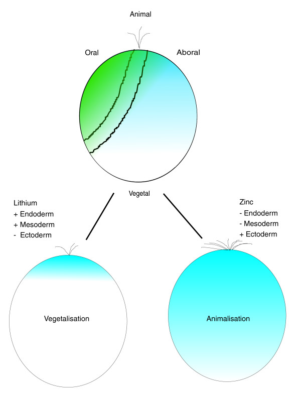
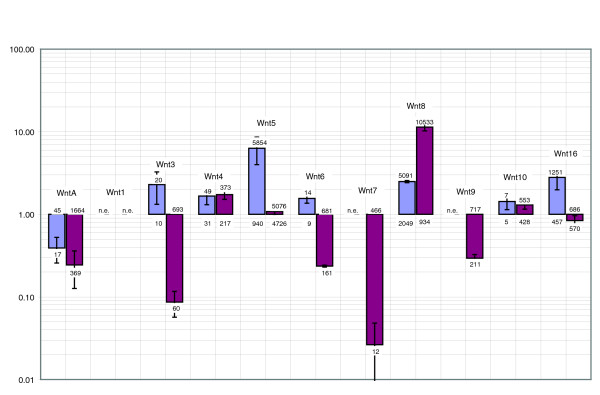
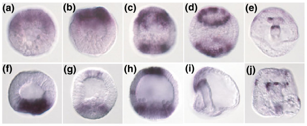
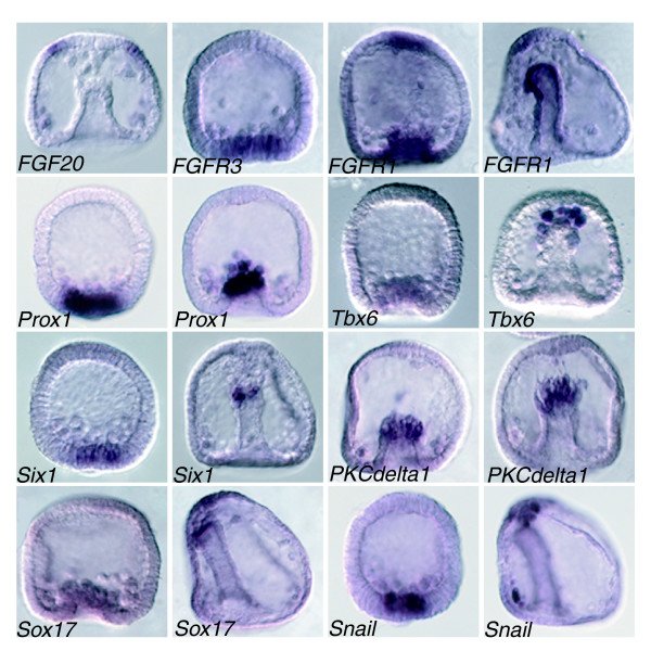
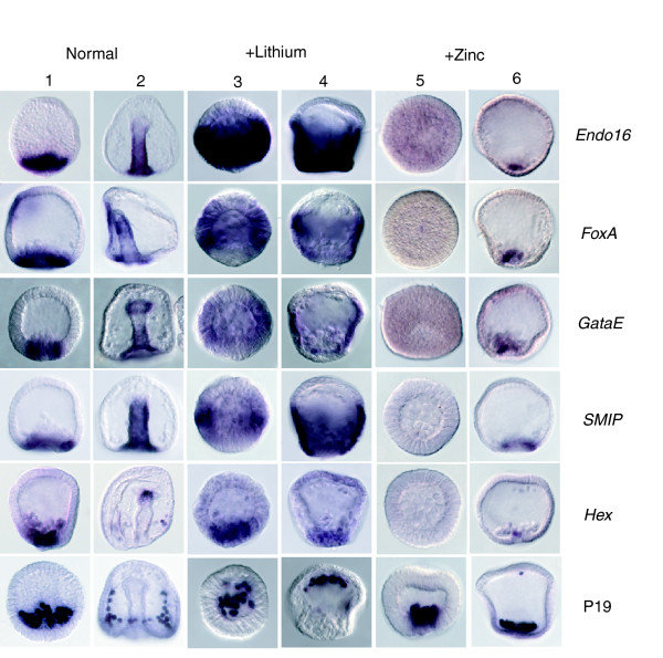
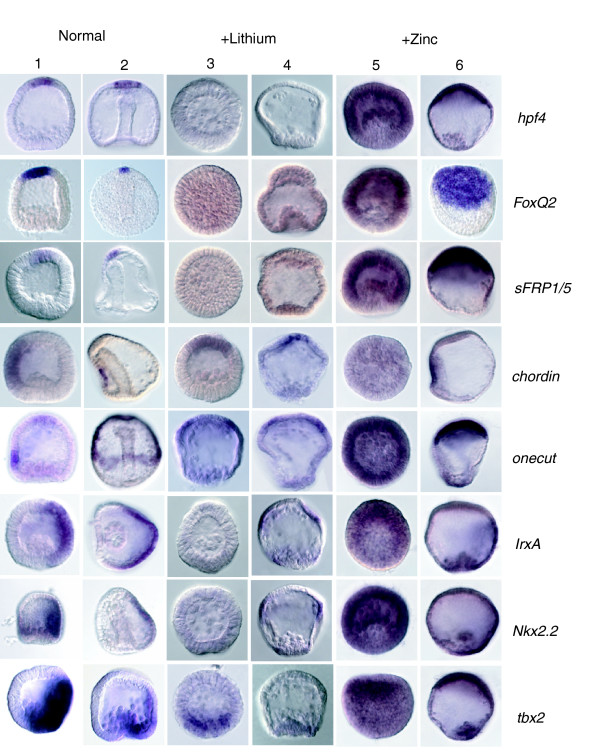
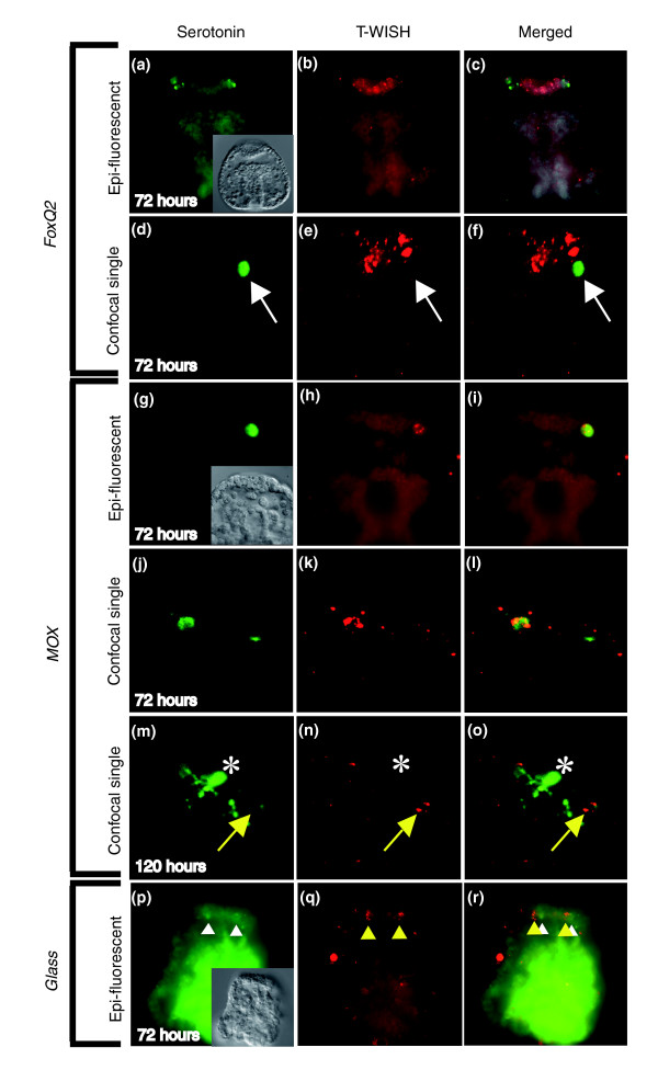
Results: Using large-scale screens on arrays carrying 50% to 70% of all genes, we identified novel territory-specific markers. Our strategy was based on computational selection of genes that are differentially expressed in lithium-treated embryos, which form excess endomesoderm, and in zinc-treated embryos, in which endomesoderm specification is blocked. Whole-mount in situ hybridization (WISH) analysis of 700 genes indicates that the apical organ region is eliminated in lithium-treated embryos. Conversely, apical and specifically neural markers are expressed more broadly in zinc-treated embryos, whereas endomesoderm signaling is severely reduced. Strikingly, the number of serotonergic neurons is amplified by at least tenfold in zinc-treated embryos. WISH analysis further indicates that there is crosstalk between the Wnt (wingless int), Notch, and fibroblast growth factor signaling pathways in secondary mesoderm cell specification and differentiation, similar to signaling cascades that function during development of presomitic mesoderm in mouse embryogenesis. We provide differential expression data for more than 4,000 genes and WISH patterns of more than 250 genes, and more than 2,400 annotated WISH images.

Conclusion: Our work provides tissue-specific expression patterns for a large fraction of the sea urchin genes that have not yet been included in existing regulatory networks and await functional integration. Furthermore, we noted neuron-inducing activity of zinc on embryonic development; this is the first observation of such activity in any organism.


Figures

Figure 1
Figure 1
Normal development and perturbations. Normal sea urchin embryos (top) develop two primary axis: the animal-vegetal axis and the oral-aboral axis. Nuclearization of β-catenin in cells on the vegetal side initiates endomesoderm specification. Later on the ectoderm is divided into an oral and aboral side, which is comparable to the dorso-ventral axis in vertebrates. Treating embryos with lithium chloride leads to enhanced nuclearization of β-catenin and, as a result, a shift in cell fate toward vegetal and formation of excess endomesoderm (left). Conversely zinc sulfate treatment prevents endomesoderm formation (right). The molecular basis for zinc sulfate action is unknown, as is the effect of these drugs on the ectoderm.
Figure 2
Figure 2
Expression of Wnt genes in lithium and zinc treated embryos. Quantitative real-time polymerase chain reaction (Q-PCR) analysis of all wingless int (Wnt) genes of the sea urchin Strongylocentrotus purpuratus. Measurements were done at blastula stage (20 hours) for lithium-treated embryos (purple bars) and gastrula stage (38 hours) for zinc-treated embryos (pink bars). Data are presented in a logarithmic style. Bars above 1 indicate upregulation and bars below 1 indicate downregulation. The numbers given on top or bottom of bars are the number of mRNA molecules/embryo in normal or treated embryos, respectively. For instance, the number of transcripts for wntA is 45 in normal 20 hours embryos and 17 in lithium-treated embryos (blue bar), and the number of transcripts of wnt5 is 940 in normal 20 hours embryos and 5,854 in lithium-treated 20 hours embryos. Where n.e. (not expressed) is indicated the gene is not expressed at this stage at all, either in control or in treated embryos. Also see Tables 1 and 3 and the text for further detail.
Figure 3
Figure 3
Opposing expression patterns of six3 and sox4. Whole-mount in situ hybridization (WISH) analysis of the developmental expression pattern of the transcription factors six3 and sox4. (a to e) six3; (f to j) sox4. The animal side is located to the top in all images. Six3 expression starts as early as 8 hours of development (8 hours embryo in panel a and 10 hours in panel b) at the animal side of the embryo. At the mesenchyme blastula stage (20 hours in panel c and flattened embryo in panel d), the animal expression clears from the central apical plate (apical organ) and at the same time forms a ring-like expression around the vegetal pole as well. In the pluteus (panel e) expression is detectable in a part of one coelomic pouch and at the forgut-midgut constriction. In contrast, sox4 is initially expressed on the vegetal side (14 hours embryo in panel f). Starting from 18 hours (panel g, and 20 hours in panel h) of development, expression also starts in the apical plate. At gastrula stage (panel i) expression is detected at the archenteron tip, and in the pluteus (panel j) expression can be detected in various secondary mesoderm cell derivatives, including some coelomic pouch cells.
Figure 4
Figure 4
Coexpression of genes in SMC cells. Whole-mount in situ hybridization (WISH) analysis of examples of signaling and transcription factor genes identified in this screen. FGF20 (Sp-FGF9/16/20), the only fibroblast growth factor present in the sea urchin genome, is expressed in primary mesenchyme cells (PMCs) and around the apical organ during gastrulation, whereas two receptors identified in this screen are expressed in adjacent secondary mesenchyme cells (SMCs; FGFR3, blastula stage) and in SMCs and the central apical region (FGFR1, left blastula, right gastrula). The transcription factors Prox1, Tbx6, Six1, Sox17, and snail are expressed in SMCs during gastrulation, as is a PKCdelta1 gene. In all pictures the animal sides of the embryos is located towards the top. Annotated images of additional stages can be found in the WISH database [22].
Figure 5
Figure 5
Expression of endomesoderm markers in normal, lithium-treated and zinc-treated embryos. Shown are whole-mount in situ hybridizations (WISHs) of endomesodermal marker genes on blastula stage (columns 1, 3, and 5) and gastrula stage (columns 2, 4, and 6) sea urchin embryos. The genes under considerations are indicated on the right hand side. Endo16, FoxA, and GataE are known, and Smip is a new gene that is expressed in the endoderm. The expression is strongly expanded in lithium-treated embryos (columns 3 and 4), whereas only at the most animal pole are ectodermal tissues left in the embryo. Blastula stage zinc-treated embryos do not exhibit any expression of endodermal markers (column 5). Gastrula stage zinc-treated embryos (column 6) do occasionally begin to express early endomesodermal markers as they recover from treatment (see Materials and methods). Hex is a transcription factor that is expressed at low levels in primary mesenchyme cells (PMCs) and predominantly in secondary mesenchyme cell (SMC) cells. Expression is upregulated in lithium-treated embryos, as determined by quantitative real-time polymerase chain reaction (Q-PCR; columns 3 and 4; compare with Table 1) but seems unchanged as determined by WISH and is eliminated in blastula stage zinc-treated embryos. P19 is a PMC-specific gene identified in the screen. Although its expression appears to be quantitatively upregulated in lithium-treated and zinc-treated embryos (see Table 2), WISH analysis indicates that the number of PMC cells forming is normal in lithium-treated or zinc-treated embryos, but that the PMCs migrate to the animal pole in lithium-treated embryos and to the vegetal pole in zinc-treated embryos. In neither case does a skeleton form.
Figure 6
Figure 6
Expression of ectoderm markers in normal, lithium-treated, and zinc-treated embryos. Shown are whole-mount in situ hybridizations (WISHs) of ectodermal marker genes on blastula stage (columns 1, 3, and 5) and gastrula stage (columns 2, 4, and 6) sea urchin embryos. The genes under considerations are indicated on the right hand side. Expression of apical plate marker genes (hpf4, FoxQ2, and secreted frizzled protein 1/5 [sFRP1/5]) is lost in lithium-treated embryos (columns 3 and 4) and expanded in zinc-treated embryos (columns 5 and 6). Expression of the oral ectoderm marker chordin is shifted to the 'new' animal pole region in lithium-treated embryos (columns 3 and 4) but lost in blastula stage zinc-treated embryos (column 5). However, ectodermal differentiation does appear to take place in zinc-treated embryos if they are left to recover for a longer period of time (column 6). The ciliated band marker gene onecut exhibits wild-type-like expression in lithium-treated embryos, with a ring of expression around the animal pole (columns 3 and 4). The apical expression domain of onecut co-expands like the other apical organ markers in zinc-treated embryos (panels 5 and 6). Strikingly, the expression of aboral ectoderm markers (IrxA, Nkx2.2, and tbx2) is lost in blastula stage lithium-treated embryos (panel 3), whereas it is enhanced in zinc-treated blastula stage embryos, in which the expression appears to be quite uniformly distributed. Tbx2 is expressed in mesodermal cells and in the aboral ectoderm in normal embryos (columns 1 and 2). Strikingly, the ectodermal expression only is lost in lithium-treated embryos, whereas the mesodermal domain remains (compare with Figure 4).
Figure 7
Figure 7
WISH analysis of Glass and Mox and immunohistochemical localization of serotonergic cells in normal and zinc-treated embryos. Whole-mount in situ hybridization (WISH) analysis identified the transcription factors (a) Glass and (b) Mox as being expressed in single cells of the apical organ. Although Glass expression is eliminated in zinc-treated embryos (b), the expression of Mox is expanded and upregulated in zinc-treated embryos (f) (also see quantitative real-time polymerase chain reaction data in Table 2). Immunohistochemical localization of serotonin in (c, d) normal and (g, h) zinc-treated embryos shows that whereas normal embryos produce four to six serotonergic cells (panel d), the number of serotonergic cells is elevated to at least 30 on average in zinc treated embryos (panel h). Panels d and h are fluorescent photographs of the same embryos depicted in transmitted light in panels c and g, respectively.
Figure 8
Figure 8
Glass, Mox, and FoxQ2 co-staining with serotonin. FoxQ2 mRNA is detected throughout the thickened neurogenic ectoderm at the animal pole of prisms (72 hours), but in 96 and 120 hour plutei there was no hybridization detectable. Tyramide amplification produces small foci of fluorescence in the cytoplasm of the cells that hybridize probe. There is diffuse background fluorescence throughout the remainder of the embryo. (a to c) In 72 hour prisms that have strong hybridization of the FoxQ2 probe to the neurogenic ectoderm, the anti-serotonin immunoreactive cells were localized outside the FoxQ2 region. (d to f) Single confocal optical section clearly shows serotonergic cells are FoxQ2 negative (white arrow). Mox mRNA was detected in the neurogenic ectoderm of prism and pluteus larvae. (g to l) In prism larvae, all of the serotonergic neurons were Mox positive. There are also some cells that are not immunoreactive with anti-serotonin, and they hybridize the Mox probe (not shown). (m to o) In plutei, neurons that are weakly immunoreactive with anti-serotonin hybridize with the Mox probe (yellow arrow). However, Mox mRNA was not detected in the neurons that strongly expressed serotonin. As the serotonergic neurons continue to differentiate during these stages, this may indicate that Mox is only expressed early in neurogenesis (asterisks in panels m to o). These preparations have relatively high background. (p to r) Glass mRNA appears not to co-localize with anti-serotonin immunoreactive cells in 72 hours prisms (white and yellow arrowheads).

Similar articles

Cited by

References

    1. Davidson EH, Rast JP, Oliveri P, Ransick A, Calestani C, Yuh CH, Minokawa T, Amore G, Hinman V, Arenas-Mena C, et al. A provisional regulatory gene network for specification of endomesoderm in the sea urchin embryo. Dev Biol. 2002;246:162–190. doi: 10.1006/dbio.2002.0635. - DOI - PubMed
    1. Davidson EH, Rast JP, Oliveri P, Ransick A, Calestani C, Yuh CH, Minokawa T, Amore G, Hinman V, Arenas-Mena C, et al. A genomic regulatory network for development. Science. 2002;295:1669–1678. doi: 10.1126/science.1069883. - DOI - PubMed
    1. Ransick A, Rast JP, Minokawa T, Calestani C, Davidson EH. New early zygotic regulators expressed in endomesoderm of sea urchin embryos discovered by differential array hybridization. Dev Biol. 2002;246:132–147. doi: 10.1006/dbio.2002.0607. - DOI - PubMed
    1. Rast JP, Cameron RA, Poustka AJ, Davidson EH. brachyury Target genes in the early sea urchin embryo isolated by differential macroarray screening. Dev Biol. 2002;246:191–208. doi: 10.1006/dbio.2002.0654. - DOI - PubMed
    1. Calestani C, Rast JP, Davidson EH. Isolation of pigment cell specific genes in the sea urchin embryo by differential macroarray screening. Development. 2003;130:4587–4596. doi: 10.1242/dev.00647. - DOI - PubMed

Publication types

LinkOut - more resources